
Ki67/MKI67 Antibody (6P3K5)
Novus Biologicals, part of Bio-Techne | Catalog # NBP3-57376
Recombinant Monoclonal Antibody

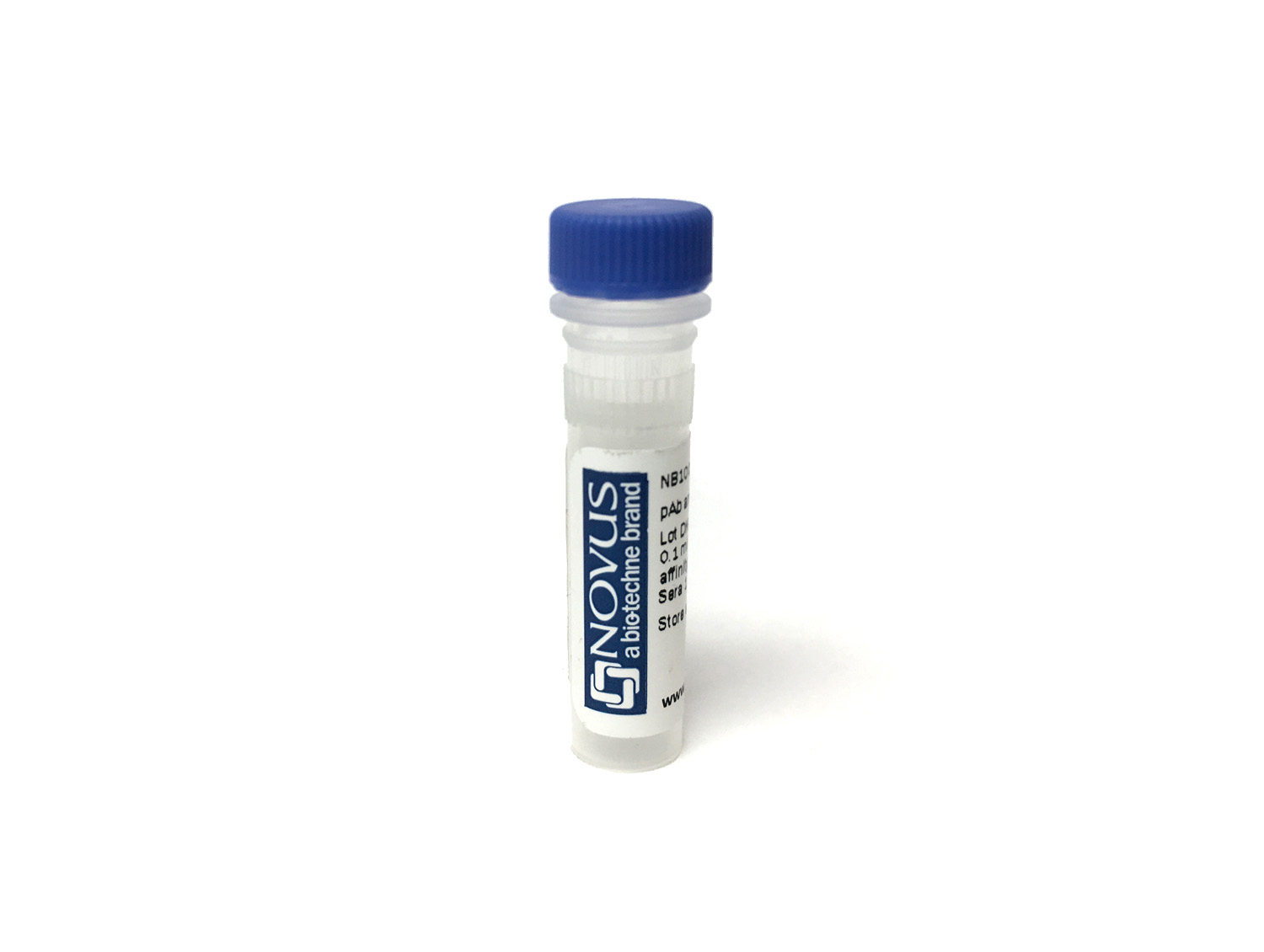
Key Product Details
Species Reactivity
Human, Mouse, Rat
Applications
Knockout Validated, Immunohistochemistry-Paraffin, Immunohistochemistry-Frozen, Western Blot, ELISA, Immunocytochemistry/ Immunofluorescence, Multiplex Immunoassay
Label
Unconjugated
Antibody Source
Recombinant Monoclonal Rabbit IgG Clone # 6P3K5
Product Specifications
Immunogen
Synthetic peptide. This information is considered to be commercially sensitive. (Swissprot# P46013)
Clonality
Monoclonal
Host
Rabbit
Isotype
IgG
Description
This antibody is highly specific and optimized for use in Immunohistochemistry.
Normal Tissues Tested
Pancreas, Lung, Tonsil, Cervix, Placenta, Colon, Kidney, Spleen, Liver, esophagus
Cancer Tissues Tested
Breast cancer, Colon cancer, Thyroid cancer, Hepatocellular Carcinoma, Hepatocholangio carcinoma, Lung adenocarcinoma, Lung squamous carcinoma
Positive Controls
Preferentially during late G1, S, G2 and M phases of the cell cycle tonsil (A moderate to strong, distinct nuclear staining reaction in 80-90% of the germinal centre B-cells in both the light and the dark zone and in the vast majority of the suprabasal squamous epithelial cells), esophagus (squamous epithelia)
Negative Controls
Liver (Hepatocytes, <1%)
Applications for Ki67/MKI67 Antibody (6P3K5)
Application
Recommended Usage
ELISA
1 ug/mL
Immunocytochemistry/ Immunofluorescence
1:50 - 1:200
Immunohistochemistry-Frozen
1:200 - 1:800
Immunohistochemistry-Paraffin
1:200 - 1:800
Multiplex Immunoassay
1:200 - 1:800
Western Blot
1:2000 - 1:10000
Application Notes
This product can be used in Multiplex Immunohistochemistry at a recommended concentration of 1:200 - 1:800.
Formulation, Preparation, and Storage
Purification
Affinity purified
Formulation
PBS, 50% glycerol, 0.05% BSA, (pH 7.3)
Preservative
0.09% Sodium Azide
Shipping
The product is shipped with polar packs. Upon receipt, store it immediately at the temperature recommended below.
Stability & Storage
Store at -20C. Avoid freeze-thaw cycles.
Background: Ki67/MKI67
Detection of Ki67 by immunostaining is commonly used as a proliferation marker in solid tumors, as well as certain hematological malignancies (3-5). The Ki67 index, which reports on positive Ki67 stained tumor cell nuclei, has been extensively studied as a prognostic biomarker in cancers such as breast cancer and cervical cancer.
References
1. Gerdes J, Schwab U, Lemke H, Stein H. (1983) Production of a mouse monoclonal antibody reactive with a human nuclear antigen associated with cell proliferation. Int J Cancer. 31:13-20. PMID: 6339421
2. Starborg M, Gell K, Brundell E and Hoog C. (1996) The murine Ki-67 cell proliferation antigen accumulates in the nucleolar and heterochromatic regions of interphase cells and at the periphery of the mitotic chromosomes in a process essential for cell cycle progression. J Cell Sci. 109:143-153. 1996
3. Karamitopoulou E, Perentes E, Tolnay M, Probst A. (1998) Prognostic significance of MIB-1, p53, and bcl-2 immunoreactivity in meningiomas. Hum Pathol. 29(2):140-5. PMID: 9490273
4. Geyer FC, Rodrigues DN, Weigelt B and Reis-Filho JS. (2012) Molecular classification of estrogen receptor-positive/luminal breast cancers. Adv Anat Pathol. 19(1):39-53. PMID: 22156833
5. Ikenberg H, Bergeron C, Schmidt D, Griesser H, Alameda F, Angeloni C, Bogers J, Dachez R, Denton K, Hariri J, Keller T, von Knebel Doeberitz M, Neumann HH, Puig-Tintore LM, Sideri M, Rehm S, Ridder R; PALMS Study Group. (2013) Screening for cervical cancer precursors with p16/Ki-67 dual-stained cytology: results of the PALMS study. J Natl Cancer Inst. 105(20):1550-7. PMID: 24096620
Long Name
Antigen Identified by Monoclonal Antibody Ki67
Alternate Names
Ki-67, KIA, MIB-1, MKI67, PPP1R105, TSG126
Gene Symbol
MKI67
Additional Ki67/MKI67 Products
Product Specific Notices for Ki67/MKI67 Antibody (6P3K5)
This product is for research use only and is not approved for use in humans or in clinical diagnosis. Primary Antibodies are guaranteed for 1 year from date of receipt.
Loading...
Loading...
Loading...
Loading...
Loading...