
Human VCAM-1/CD106 Antibody
R&D Systems, part of Bio-Techne | Catalog # BBA19

Scientific images may be resized, cropped, or adjusted for brightness or contrast for presentation purposes. These updates are limited to presentation and do not affect the underlying scientific interpretation of the data. Where available, additional source data may be provided upon request.
Key Product Details
Species Reactivity
Validated:
Human
Cited:
Human, Mouse, Rabbit
Applications
Validated:
Immunohistochemistry, Western Blot, Simple Western
Cited:
Immunohistochemistry, Immunohistochemistry-Paraffin, Western Blot, Immunocytochemistry
Label
Unconjugated
Antibody Source
Polyclonal Goat Serum
Product Specifications
Immunogen
Chinese hamster ovary cell line CHO-derived recombinant human VCAM-1
Specificity
Detects VCAM‑1/CD106 in Western blots. VCAM‑1/CD106 has been screened using CHO cells transfected with cDNAs for ICAM-1 and VCAM-1.
Clonality
Polyclonal
Host
Goat
Isotype
Serum
Scientific Data Images for Human VCAM-1/CD106 Antibody
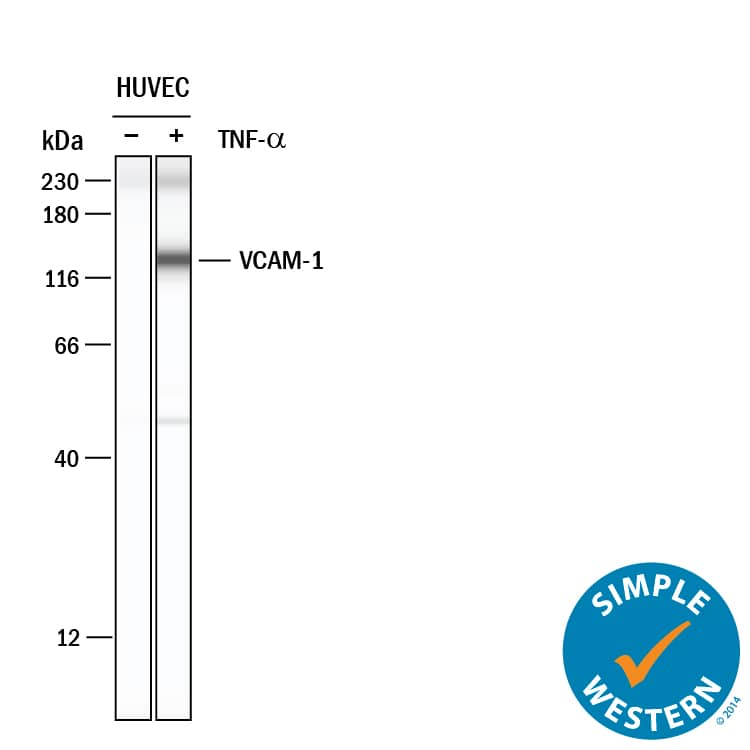
Detection of Human VCAM-1/CD106 by Simple WesternTM.
Simple Western lane view shows lysates of HUVEC human umbilical vein endothelial cells untreated (-) or treated (+) with 10 ng/ml Recombinant Human TNF-alpha (210-TA) for 24 hrs, loaded at 0.2 mg/mL. A specific band was detected for VCAM-1/CD106 at approximately 132 kDa (as indicated) using 20 µg/mL of Goat Anti-Human VCAM-1/CD106 Polyclonal Antibody (Catalog # BBA19) . This experiment was conducted under reducing conditions and using the 12-230 kDa separation system.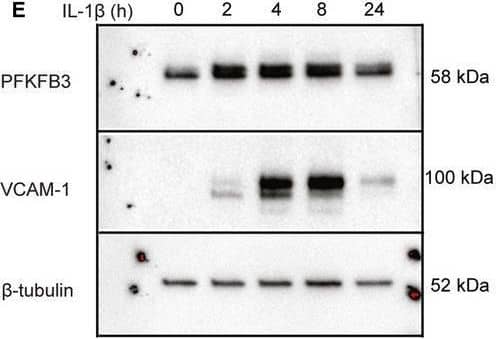
Detection of VCAM-1/CD106 by Western Blot
IL‐1 beta stimulation increases endothelial glucose uptake, glycolysis, and PFKFB3 expression. (A) Uptake of 14C‐2‐deoxy‐d‐glucose in IL‐1 beta‐stimulated HUVECs (1 ng·mL−1, 16 h; mean ± SD; n = 3 donors). (B) Rate of glycolysis in IL‐1 beta‐stimulated HUVECs (n = 3 donors). (C) Glycolysis fold change 6 h after stimulation with different doses of IL‐1 beta (n = 3 donors). (D) RT‐qPCR of PFKFB3 following in IL‐1 beta‐stimulated HUVECs (1 ng·mL−1) (n = 3 donors). (E) Representative immunoblot for PFKFB3 (top), VCAM‐1 (middle) and beta‐tubulin loading control (bottom) in IL‐1 beta‐stimulated HUVECs (1 ng·mL−1, 2–24 h). (F) Densitometric quantification of PFKFB3 relative to beta‐tubulin loading control (n = 5 donors). (G) RT‐qPCR of VCAM‐1 in IL‐1 beta‐stimulated HUVECs (1 ng·mL−1, 2–24 h) (n = 3 donors). (H) Densitometric quantification of VCAM‐1 relative to beta‐tubulin loading control (n = 5 donors). B–D and F–H: lines show means, and dots show individual donors. *P < 0.05, **P < 0.01, ***P < 0.001, ****P < 0.0001. Image collected and cropped by CiteAb from the following open publication (https://pubmed.ncbi.nlm.nih.gov/33979025), licensed under a CC-BY license. Not internally tested by R&D Systems.Applications for Human VCAM-1/CD106 Antibody
Application
Recommended Usage
Immunohistochemistry
1:200 dilution
Sample: Immersion fixed paraffin-embedded sections of human kidney subjected to Antigen Retrieval Reagent-Basic (Catalog # CTS013)
Sample: Immersion fixed paraffin-embedded sections of human kidney subjected to Antigen Retrieval Reagent-Basic (Catalog # CTS013)
Simple Western
20 µg/mL
Sample: HUVEC human umbilical vein endothelial cells
Sample: HUVEC human umbilical vein endothelial cells
Western Blot
1:1000 dilution
Sample: Recombinant Human VCAM-1/CD106 (Catalog # 809-VR)
Sample: Recombinant Human VCAM-1/CD106 (Catalog # 809-VR)
Reviewed Applications
Read 1 review rated 5 using BBA19 in the following applications:
Formulation, Preparation, and Storage
Reconstitution
Reconstitute in 0.5 mL of sterile water.
Formulation
Lyophilized from a 0.2 μm filtered solution in Serum.
Shipping
The product is shipped at ambient temperature. Upon receipt, store it immediately at the temperature recommended below.
Stability & Storage
Use a manual defrost freezer and avoid repeated freeze-thaw cycles.
- 12 months from date of receipt, -20 to -70 °C as supplied.
- 1 month, 2 to 8 °C under sterile conditions after reconstitution.
- 6 months, -20 to -70 °C under sterile conditions after reconstitution.
Background: VCAM-1/CD106
Vascular cell adhesion molecule (VCAM-1) is expressed on the surface of activated endothelial cells and macrophages. It binds to leukocyte integrins VLA-4 and alpha4 beta7 to promote leukocyte adhesion at sites of inflamed vasculature.
Long Name
Vascular Cell Adhesion Molecule 1
Alternate Names
CD106, VCAM1
Gene Symbol
VCAM1
Additional VCAM-1/CD106 Products
Product Documents for Human VCAM-1/CD106 Antibody
Product Specific Notices for Human VCAM-1/CD106 Antibody
For research use only
Loading...
Loading...
Loading...
Loading...
Loading...