
Human TGF-beta RII Fluorescein-conjugated Antibody
R&D Systems, part of Bio-Techne | Catalog # FAB241F

Scientific images may be resized, cropped, or adjusted for brightness or contrast for presentation purposes. These updates are limited to presentation and do not affect the underlying scientific interpretation of the data. Where available, additional source data may be provided upon request.
Key Product Details
Species Reactivity
Validated:
Human
Cited:
Human, Mouse
Applications
Validated:
Flow Cytometry
Cited:
Flow Cytometry, FACS
Label
FITC (Excitation = 488 nm, Emission = 515-545 nm)
Antibody Source
Monoclonal Mouse IgG1 Clone # 25508
Product Specifications
Immunogen
Mouse myeloma cell line NS0-derived recombinant human TGF-beta RII
Ile24-Asp159
Accession # P37173.2
Ile24-Asp159
Accession # P37173.2
Specificity
This antibody has been validated by flow cytometry on TGF-beta RII-transfected cells only. In contrast to Catalog # FAB2411F, minimal staining of PBMC lymphocytes was observed with this antibody.
Clonality
Monoclonal
Host
Mouse
Isotype
IgG1
Scientific Data Images for Human TGF-beta RII Fluorescein-conjugated Antibody
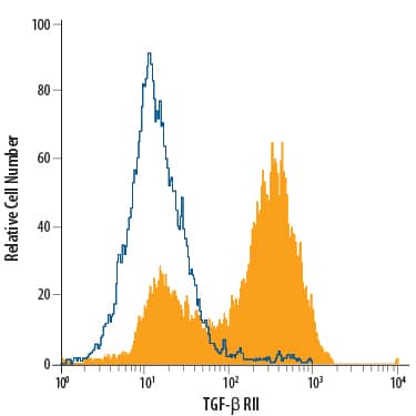
Detection of TGF-beta RII in NS0 Mouse Cell Line Transfected with TGF-beta RII by Flow Cytometry.
NS0 mouse myeloma cell line transfected with TGF-beta RII was stained with Mouse Anti-Human TGF-beta RII Fluorescein-conjugated Monoclonal Antibody (Catalog # FAB241F, filled histogram) or isotype control antibody (Catalog # IC002F, open histogram). View our protocol for Staining Membrane-associated Proteins.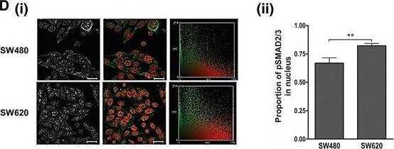
Detection of Human TGF-beta RII by Immunocytochemistry/Immunofluorescence
Investigating the status of the canonical TGF-beta pathway and levels of Hsp90 in the genetically paired SW480 primary and SW620 secondary tumour-derived colon cancer cell lines. a The secretion of TGF-beta 1 by SW480 and SW620 cells was determined by means of an ELISA to quantify the levels of extracellular TGF-beta 1 in spent culture medium for a given cell number after 12 h incubation. Data represent the average of three independent experiments performed in triplicate and error bars indicate the standard error in the mean. b The levels of TGF-beta RII receptor on the cell surface was determined by flow cytometry using a fluorescein isothiocyanate-conjugated antibody. Data was analysed using FlowJo software and gating carried out according to the IgG1 isotype control. Histograms show a shift in fluorescence for each sample (black line, unshaded) compared to the isotype control (grey shading) and the percentage of positive events is indicated for each sample. Data are representative of three independent experiments showing consistent results. c Intracellular levels of TGF-beta 1 in SW480 and SW620 cells were determined by (i) western blot analysis and compared to a histone loading control. The images are cropped from the full length western blot provided as Additional file 1. (ii) The normalized levels of pre-pro TGF beta1 from the western blot in (i) were determined by densitometry using ImageJ and calculated according the histone loading control. AU: arbitrary units. The positive control represents commercially obtained recombinant human TGF beta1 in its active form. d Activation of the TGF-beta canonical pathway was determined by the phosphorylation and nuclear localization of Smad2/3. SW480 and SW620 cells were stained for pSMAD2/3 and nuclei were stained with Hoescht-33342. Immunofluorescence was detected using the Zeiss LSM 510 Meta laser scanning confocal microscope and the images analysed using Axiovision LE 4.7.1 software (Zeiss). (i) The first column shows the level of pSmad2/3 stainiApplications for Human TGF-beta RII Fluorescein-conjugated Antibody
Application
Recommended Usage
Flow Cytometry
10 µL/106 cells
Sample: NS0 mouse myeloma cell line transfected with TGF‑ beta RII
Sample: NS0 mouse myeloma cell line transfected with TGF‑ beta RII
Reviewed Applications
Read 1 review rated 4 using FAB241F in the following applications:
Spectra Viewer
Plan Your Experiments
Use our spectra viewer to interactively plan your experiments, assessing multiplexing options. View the excitation and emission spectra for our fluorescent dye range and other commonly used dyes.
Spectra Viewer
Formulation, Preparation, and Storage
Purification
Protein A or G purified from hybridoma culture supernatant
Formulation
Supplied in a saline solution containing BSA and Sodium Azide.
Shipping
The product is shipped with polar packs. Upon receipt, store it immediately at the temperature recommended below.
Stability & Storage
Protect from light. Do not freeze.
- 12 months from date of receipt, 2 to 8 °C as supplied.
Background: TGF-beta RII
TGF-beta RII is a membrane bound serine/threonine kinase. Upon ligand binding, TGF-beta RII interacts with TGF-beta RI to form the heteromeric signaling complex that transduces TGF-beta signals. A splice variant of the type II receptor, TGF-beta RIIb, containing a 25 amino acid residue insertion near the N-terminus of the mature protein has also been described.
Long Name
Transforming Growth Factor beta Receptor II
Alternate Names
TGF-bRII, TGFbetaRII, TGFBR2
Gene Symbol
TGFBR2
UniProt
Additional TGF-beta RII Products
Product Documents for Human TGF-beta RII Fluorescein-conjugated Antibody
Product Specific Notices for Human TGF-beta RII Fluorescein-conjugated Antibody
For research use only
Loading...
Loading...
Loading...
Loading...
Loading...