
Human/Mouse/Rat Semaphorin 5A Antibody
R&D Systems, part of Bio-Techne | Catalog # AF5896

Discontinued Product
AF5896 has been discontinued.
View all Semaphorin 5A products.
Key Product Details
Species Reactivity
Validated:
Human, Mouse, Rat
Cited:
Human, Mouse, Transgenic Mouse
Applications
Validated:
Immunohistochemistry, Western Blot, Immunocytochemistry
Cited:
Immunohistochemistry, Western Blot, Flow Cytometry
Label
Unconjugated
Antibody Source
Polyclonal Sheep IgG
Product Specifications
Immunogen
Chinese hamster ovary cell line CHO-derived recombinant human Semaphorin 5A
Glu23-Thr765
Accession # AAC09473
Glu23-Thr765
Accession # AAC09473
Specificity
Detects human, mouse, and rat Semaphorin 5A in direct ELISAs and Western blots. In direct ELISAs, less than 1% cross-reactivity with recombinant human (rh) Sema3B, rhSema4C, and rhSema6A is observed.
Clonality
Polyclonal
Host
Sheep
Isotype
IgG
Scientific Data Images for Human/Mouse/Rat Semaphorin 5A Antibody
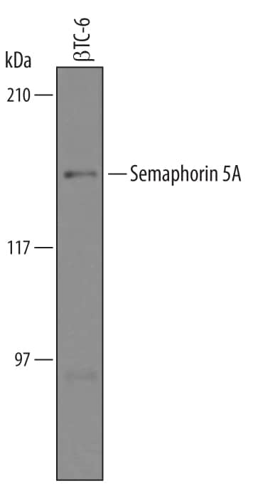
Detection of Mouse Semaphorin 5A by Western Blot.
Western blot shows lysates of betaTC-6 mouse beta cell insulinoma cell line. PVDF Membrane was probed with 1 µg/mL of Sheep Anti-Human/Mouse/Rat Semaphorin 5A Antigen Affinity-purified Polyclonal Antibody (Catalog # AF5896) followed by HRP-conjugated Anti-Sheep IgG Secondary Antibody (Catalog # HAF016). A specific band was detected for Semaphorin 5A at approximately 135 kDa (as indicated). This experiment was conducted under reducing conditions and using Immunoblot Buffer Group 8.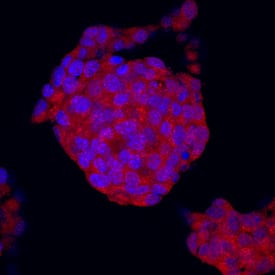
Semaphorin 5A in betaTC‑6 Mouse Cell Line.
Semaphorin 5A was detected in immersion fixed betaTC-6 mouse beta cell insulinoma cell line using Sheep Anti-Human/Mouse/Rat Semaphorin 5A Antigen Affinity-purified Polyclonal Antibody (Catalog # AF5896) at 10 µg/mL for 3 hours at room temperature. Cells were stained using the NorthernLights™ 557-conjugated Anti-Sheep IgG Secondary Antibody (red; Catalog # NL010) and counterstained with DAPI (blue). Specific staining was localized to cytoplasm. View our protocol for Fluorescent ICC Staining of Cells on Coverslips.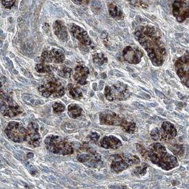
Semaphorin 5A in Human Pancreas.
Semaphorin 5A was detected in immersion fixed paraffin-embedded sections of human pancreas using Sheep Anti-Human/Mouse/Rat Semaphorin 5A Antigen Affinity-purified Polyclonal Antibody (Catalog # AF5896) at 5 µg/mL overnight at 4 °C. Tissue was stained using the Anti-Sheep HRP-DAB Cell & Tissue Staining Kit (brown; Catalog # CTS019) and counterstained with hemotoxylin (blue). Specific staining was localized to the plasma membranes of epithelial cells. View our protocol for Chromogenic IHC Staining of Paraffin-embedded Tissue Sections.Applications for Human/Mouse/Rat Semaphorin 5A Antibody
Application
Recommended Usage
Immunocytochemistry
5-15 µg/mL
Sample: Immersion fixed betaTC‑6 mouse beta cell insulinoma cell line
Sample: Immersion fixed betaTC‑6 mouse beta cell insulinoma cell line
Immunohistochemistry
5-15 µg/mL
Sample: Immersion fixed paraffin-embedded sections of human pancreas
Sample: Immersion fixed paraffin-embedded sections of human pancreas
Western Blot
1 µg/mL
Sample: betaTC‑6 mouse beta cell insulinoma cell line
Sample: betaTC‑6 mouse beta cell insulinoma cell line
Reviewed Applications
Read 1 review rated 5 using AF5896 in the following applications:
Formulation, Preparation, and Storage
Purification
Antigen Affinity-purified
Reconstitution
Reconstitute at 0.2 mg/mL in sterile PBS. For liquid material, refer to CoA for concentration.
Formulation
Lyophilized from a 0.2 μm filtered solution in PBS with Trehalose. *Small pack size (SP) is supplied either lyophilized or as a 0.2 µm filtered solution in PBS.
Shipping
Lyophilized product is shipped at ambient temperature. Liquid small pack size (-SP) is shipped with polar packs. Upon receipt, store immediately at the temperature recommended below.
Stability & Storage
Use a manual defrost freezer and avoid repeated freeze-thaw cycles.
- 12 months from date of receipt, -20 to -70 °C as supplied.
- 1 month, 2 to 8 °C under sterile conditions after reconstitution.
- 6 months, -20 to -70 °C under sterile conditions after reconstitution.
Background: Semaphorin 5A
Semaphorin 5A (Sema5A, previously called SemaF) is a 140 kDa protein of the semaphorin family of axon guidance molecules (1‑4). Class 5 semaphorins are type I transmembrane glycoproteins with an N-terminal Sema domain and multiple juxtamembrane type 1 thrombospondin (TSP) repeats within their extracellular domains (1‑3). Sema5A is expressed developmentally in oligodendrocytes, neuroepithelial cells surrounding retinal axons, the base of limb buds, the cardiac atrial septum and endocardial cushions, and the mesoderm surrounding cranial vessels (4‑6). The human Sema5A cDNA encodes a 22 amino acid (aa) signal sequence, a 946 aa extracellular domain (ECD), a 22 aa transmembrane sequence and an 85 aa cytoplasmic portion. Within aa 23‑765, which includes the sema domain and four of the seven TSP repeats, human Sema5A shares 93% aa identity with corresponding mouse, rat, and canine sequences. Semaphorins typically transduce signals through transmembrane plexins (1, 2). The sema domain of Sema5A binds plexin B3, triggering signaling via HGF R/c-Met (7). Both Sema5A and plexin B3 are expressed postnatally during differentiation and migration of central nervous system oligodendrocytes. However, plexin B3 is not significantly expressed prenatally and therefore unlikely to be the Sema5A receptor during development (7, 8). The Sema5A TSP repeats interact with either heparin sulfate or chondroitin sulfate proteoglycans (HSPG, CSPG) (9). HSPG interaction promotes attraction, while CSPG interaction promotes repulsion and is essential for axon fasciculation, independent of plexin B3 (9, 10). Sema5A mutations have been implicated in the genetic syndrome, cri-du-chat, while some polymorphisms may increase risk for neurodegenerative diseases such as Parkinson’s (3, 11). Sema5A expression may be upregulated in metastatic cancer cells and downregulated in autism (12, 13).
References
- Flannery, E. and M. Duman-Scheel (2009) Curr. Drug Targets 10:611.
- Zhou, Y. et al. (2008) Trends Biol. Sci. 33:161.
- Simmons, A.D. et al. (1998) Biochem. Biophys. Res. Commun. 242:685.
- Oster, S.F. et al. (2003) Development 130:775.
- Goldberg, J.L. et al. (2004) J. Neurosci. 24:4989.
- Fiore, R. et al. (2005) Mol. Cell. Biol. 25:2310.
- Artigiani, S. et al. (2004) EMBO Rep. 5:710.
- Worzfeld, T. et al. (2004) Eur. J. Neurosci. 19:2622.
- Kantor, D.B. et al. (2004) Neuron 44:961.
- Hilario, J.D. et al. (2008) Dev. Biol. 326:190.
- Lin, L. et al. (2009) Trends Neurosci. 32:142.
- Pan, G-Q. et al. (2009) World J. Gastroenterol. 15:2800.
- Melin, M. et al. (2006) Neuropsychobiology 54:64.
Alternate Names
SEMA5A, Semaphorin-F
Gene Symbol
SEMA5A
UniProt
Additional Semaphorin 5A Products
Product Documents for Human/Mouse/Rat Semaphorin 5A Antibody
Product Specific Notices for Human/Mouse/Rat Semaphorin 5A Antibody
For research use only
Loading...
Loading...
Loading...
Loading...
Loading...