
Human DISP1 Antibody
R&D Systems, part of Bio-Techne | Catalog # AF3549

Scientific images may be resized, cropped, or adjusted for brightness or contrast for presentation purposes. These updates are limited to presentation and do not affect the underlying scientific interpretation of the data. Where available, additional source data may be provided upon request.
Conjugate
Catalog #
Key Product Details
Species Reactivity
Human
Applications
Western Blot, Immunocytochemistry
Label
Unconjugated
Antibody Source
Polyclonal Sheep IgG
Product Specifications
Immunogen
E. coli-derived recombinant human DISP1
Glu212-Lys492, Ser746-Thr984
Accession # NP_116279
Glu212-Lys492, Ser746-Thr984
Accession # NP_116279
Specificity
Detects human DISP1 in direct ELISAs and Western blots.
Clonality
Polyclonal
Host
Sheep
Isotype
IgG
Scientific Data Images for Human DISP1 Antibody
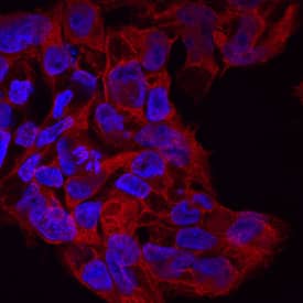
DISP1 in BG01V Human Embryonic Stem Cells.
DISP1 was detected in immersion fixed BG01V human embryonic stem cells using Sheep Anti-Human DISP1 Antigen Affinity-purified Polyclonal Antibody (Catalog # AF3549) at 10 µg/mL for 3 hours at room temperature. Cells were stained using the NorthernLights™ 557-conjugated Anti-Sheep IgG Secondary Antibody (red; Catalog # NL010) and counterstained with DAPI (blue). Specific staining was localized to cytoplasm. View our protocol for Fluorescent ICC Staining of Cells on Coverslips.
Detection of DISP1 by Western Blot
Impaired Shh release from Disp−/− cells. (A) Alignment of targeted disp gene sequences from Disp−/− cells and from non-targeted (nt Ctrl) cells. (A′) Schematic representation of the Disp protein structure. An asterisk indicates the CRISPR/Cas9-generated stop codon introduced at position 323, deleting 11 of 12 TM domains that together represent ∼80% of the protein sequence. L1 and L2 indicate extracellular loops. TM2–TM6 (colored red) constitute the SSD. (B,C) Immunoblots of cellular (c) and released (into the medium, m) Shh (B) and unlipidated control C25SShhN (C) in nt Ctrl and Disp−/− cells in the presence of Scube2. Arrows indicate solubilized Shh and the arrowhead indicates accumulated cellular material in Disp−/− cells. (D) In the absence of Scube2, Shh processing into serum-free medium was abolished in nt Ctrl and Disp−/− cells. Instead, both cell types released similar amounts of unprocessed protein. In B,C,D, anti-beta -actin blots ( alpha beta-actin) and Ponceau S staining of residual serum albumin (PonS) serve as loading controls. (B′,C′,D′) Quantifications of relative Shh (B′,D′) and C25SShhN (C′) release from nt Ctrl and Disp−/− cells. Ratios of solubilized versus cellular Shh were determined and expressed relative to Shh release from nt Ctrl cells (black bars). Data are mean±s.d. n=21 in B′, n=8 in C′ and n=5 in D′. ****P<0.0001; ns, not significant (two-tailed unpaired t-test). Image collected and cropped by CiteAb from the following open publication (https://pubmed.ncbi.nlm.nih.gov/34308968), licensed under a CC-BY license. Not internally tested by R&D Systems.Applications for Human DISP1 Antibody
Application
Recommended Usage
Immunocytochemistry
5-15 µg/mL
Sample: Immersion fixed BG01V human embryonic stem cells
Sample: Immersion fixed BG01V human embryonic stem cells
Western Blot
0.1 µg/mL
Sample: Recombinant Human DISP1
Sample: Recombinant Human DISP1
Formulation, Preparation, and Storage
Purification
Antigen Affinity-purified
Reconstitution
Reconstitute at 0.2 mg/mL in sterile PBS. For liquid material, refer to CoA for concentration.
Formulation
Lyophilized from a 0.2 μm filtered solution in PBS with Trehalose. *Small pack size (SP) is supplied either lyophilized or as a 0.2 µm filtered solution in PBS.
Shipping
Lyophilized product is shipped at ambient temperature. Liquid small pack size (-SP) is shipped with polar packs. Upon receipt, store immediately at the temperature recommended below.
Stability & Storage
Use a manual defrost freezer and avoid repeated freeze-thaw cycles.
- 12 months from date of receipt, -20 to -70 °C as supplied.
- 1 month, 2 to 8 °C under sterile conditions after reconstitution.
- 6 months, -20 to -70 °C under sterile conditions after reconstitution.
Background: DISP1
DISP1 is a multispan (12) transmembrane protein with cytoplasmic N- and C-terminal tails. It contains a stero/sensing domain (SSD) and has been shown to play a role in regulating the release of cholesterol-modified SHH. Within the regions used for immunization, human and mouse DISP1 share 93% amino acid sequence homology.
Long Name
Dispatched 1
Alternate Names
DISPA, Dispatched
Gene Symbol
DISP1
UniProt
Additional DISP1 Products
Product Documents for Human DISP1 Antibody
Product Specific Notices for Human DISP1 Antibody
For research use only
Loading...
Loading...
Loading...
Loading...
Loading...