
Human CXCL10/IP‑10/CRG‑2 Biotinylated Antibody
R&D Systems, part of Bio-Techne | Catalog # BAF266

Scientific images may be resized, cropped, or adjusted for brightness or contrast for presentation purposes. These updates are limited to presentation and do not affect the underlying scientific interpretation of the data. Where available, additional source data may be provided upon request.
Conjugate
Catalog #
Key Product Details
Species Reactivity
Validated:
Human
Cited:
Human
Applications
Validated:
ELISA Detection (Matched Antibody Pair), Intracellular Staining by Flow Cytometry
Cited:
Immunocytochemistry, ELISA Detection, ELISA Development, ELISA Development (Detection), Luminex Development
Label
Biotin
Antibody Source
Polyclonal Goat IgG
Product Specifications
Immunogen
E. coli-derived recombinant human CXCL10/IP-10
Specificity
Detects human CXCL10/IP-10/CRG-2 in ELISAs and Western blots. In sandwich ELISAs, less than 0.1% cross-reactivity with recombinant mouse CRG‑2, recombinant human (rh) Lymphotactin, rhGRO alpha, and rhMIG is observed.
Clonality
Polyclonal
Host
Goat
Isotype
IgG
Scientific Data Images for Human CXCL10/IP‑10/CRG‑2 Biotinylated Antibody
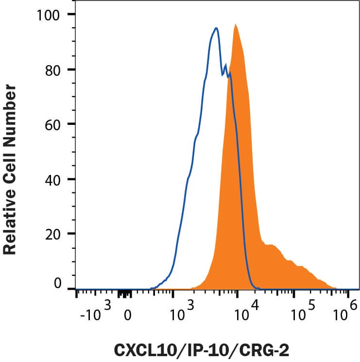
Detection of CXCL10/IP-10/CRG02 in Human PBMC by Flow Cytometry.
Human PBMC treated (filled histogram) and untreated (open histogram) with 75 ng/mL rhIFNy and monensin overnight were stained with Goat anti-human CXCL10/IP-10/CRG02 biotin (BAF266) followed by Phycoerythrin-conjugated Streptavidin (F0040). To facilitate intracellular staining, cells were fixed with Flow Cytometry Fixation Buffer (FC004) and permeabilized with Flow Cytometry Permeabilization/Wash Buffer I (FC005). View our protocol for Staining Intracellular Molecules.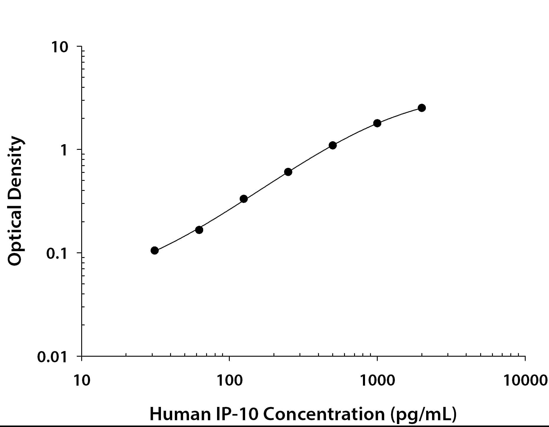
Human CXCL10 / IP-10 / CRG-2 ELISA Standard Curve
Recombinant Human CXCL10/IP-10/CRG-2 (Catalog # 266-IP) was serially diluted and captured by Mouse Anti-Human CXCL10/IP-10/CRG-2 Monoclonal Antibody (Catalog # MAB266) coated on a Clear Polystyrene Microplate (Catalog # DY990). Goat Anti-Human CXCL10/IP-10/CRG-2 Antigen Affinity-purified Polyclonal Antibody (Catalog # AF-266-NA) was biotinylated and incubated with the protein captured on the plate. Detection of the standard curve was achieved by incubating Streptavidin-HRP (Catalog # DY998)Applications for Human CXCL10/IP‑10/CRG‑2 Biotinylated Antibody
Application
Recommended Usage
Intracellular Staining by Flow Cytometry
2.5 µg/106 cells
Sample: Human peripheral blood monocytes treated with Recombinant Human IFN‑ gamma (Catalog # 285‑IF), fixed with paraformaldehyde, and permeabilized with saponin
Sample: Human peripheral blood monocytes treated with Recombinant Human IFN‑ gamma (Catalog # 285‑IF), fixed with paraformaldehyde, and permeabilized with saponin
Human CXCL10/IP-10 Sandwich Immunoassay
ELISA Detection (Matched Antibody Pair)
Please Note: Optimal dilutions of this antibody should be experimentally determined.
Reviewed Applications
Read 2 reviews rated 4.5 using BAF266 in the following applications:
Formulation, Preparation, and Storage
Purification
Antigen Affinity-purified
Reconstitution
Reconstitute at 0.2 mg/mL in sterile PBS.
Formulation
Lyophilized from a 0.2 μm filtered solution in PBS with BSA as a carrier protein.
Shipping
The product is shipped at ambient temperature. Upon receipt, store it immediately at the temperature recommended below.
Stability & Storage
Use a manual defrost freezer and avoid repeated freeze-thaw cycles.
- 12 months from date of receipt, -20 to -70 °C as supplied.
- 1 month, 2 to 8 °C under sterile conditions after reconstitution.
- 6 months, -20 to -70 °C under sterile conditions after reconstitution.
Background: CXCL10/IP-10/CRG-2
CXCL10 was originally identified as an IFN-gamma -inducible gene in monocytes, fibroblasts, and endothelial cells. It has since been shown that CXCL10 mRNA is also induced by LPS, IL-1 beta, TNF-alpha, IL-12, and viruses. Additional cell types that have been shown to express CXCL10 include activated T-lymphocytes, splenocytes, keratinocytes, osteoblasts, astrocytes, and smooth muscle cells. CXCL10 is also expressed in psoriatic and lepromatous lesions of skin. The mouse homologue of human CXCL10, CRG-2, has been cloned and shown to share approximately 67% amino acid (aa) sequence identity with human CXCL10. Human CXCL10 cDNA encodes a 98 aa residue precursor protein with a 21 aa residue signal peptide that is cleaved to form the 77 aa residue secreted protein. The amino acid sequence of CXCL10 identified the protein as a member of the chemokine alpha subfamily that lacks the ELR domain. CXCL10 has been shown to be a chemoattractant for activated T-lymphocytes. CXCL10 has been reported to be a potent inhibitor of angiogenesis and to display a potent thymus-dependent anti-tumor effect. A chemokine receptor specific for CXCL10 and MIG has been cloned and shown to be highly expressed in IL-2-activated T-lymphocytes.
References
- Loetscher, M. et al. (1996) J. Exp. Med. 184:963.
- Wang, X. et al. (1996) J. Biol. Chem. 271:24286.
Alternate Names
CRG-2, CRG2, IP-10
Gene Symbol
CXCL10
Additional CXCL10/IP-10/CRG-2 Products
Product Documents for Human CXCL10/IP‑10/CRG‑2 Biotinylated Antibody
Product Specific Notices for Human CXCL10/IP‑10/CRG‑2 Biotinylated Antibody
For research use only
Loading...
Loading...
Loading...
Loading...
Loading...
Loading...