Human Coagulation Factor III/Tissue Factor Antibody
R&D Systems, part of Bio-Techne | Catalog # MAB2339


Key Product Details
Validated by
Species Reactivity
Validated:
Cited:
Applications
Validated:
Cited:
Label
Antibody Source
Product Specifications
Immunogen
Gly34-Glu251
Accession # P13726
Specificity
Clonality
Host
Isotype
Scientific Data Images for Human Coagulation Factor III/Tissue Factor Antibody
Detection of Human Coagulation Factor III by Western Blot.
Western blot shows lysates of THP-1 human acute monocytic leukemia cell line treated (+) with TNF-a. PVDF membrane was probed with 2 µg/mL of Mouse Anti-Human Coagulation Factor III/Tissue Factor Monoclonal Antibody (Catalog # MAB2339) followed by HRP-conjugated Anti-Mouse IgG Secondary Antibody (Catalog # HAF007). A specific band was detected for Coagulation Factor III at approximately 50 and 54 kDa (as indicated). This experiment was conducted under reducing conditions and using Immunoblot Buffer Group 1.Detection of Human Coagulation Factor III/Tissue Factor by Western Blot.
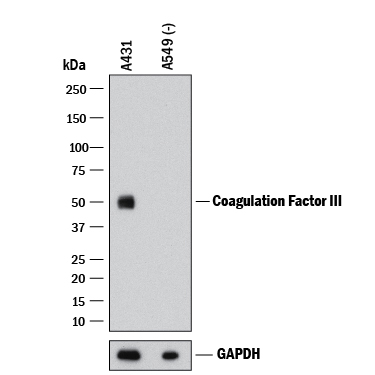
Western blot shows lysates of A431 human epithelial carcinoma cell line and A549 human lung carcinoma cell line (negative control). PVDF membrane was probed with 1 µg/mL of Mouse Anti-Human Coagulation Factor III/Tissue Factor Monoclonal Antibody (Catalog # MAB2339) followed by HRP-conjugated Anti-Mouse IgG Secondary Antibody (HAF018). A specific band was detected for Coagulation Factor III/Tissue Factor at approximately 48 kDa (as indicated). GAPDH (MAB5718) is shown as a loading control. This experiment was conducted under reducing conditions and using Western Blot Buffer Group 1.Applications for Human Coagulation Factor III/Tissue Factor Antibody
Immunoprecipitation
Sample: Conditioned cell culture medium spiked with Recombinant Human Coagulation Factor III/Tissue Factor (Catalog # 2339-PA), see our available Western blot detection antibodies
Western Blot
Sample: THP‑1 human acute monocytic leukemia cell line treated with TNF-alpha and A431 human epithelial carcinoma cell line
Human Coagulation Factor III/Tissue Factor Sandwich Immunoassay
Reviewed Applications
Read 2 reviews rated 5 using MAB2339 in the following applications:
Formulation, Preparation, and Storage
Purification
Reconstitution
Formulation
Shipping
Stability & Storage
- 12 months from date of receipt, -20 to -70 °C as supplied.
- 1 month, 2 to 8 °C under sterile conditions after reconstitution.
- 6 months, -20 to -70 °C under sterile conditions after reconstitution.
Background: Coagulation Factor III/Tissue Factor
Coagulation Factor III/Tissue Factor (TF), also known as thromboplastin and CD142, is a type I transmembrane protein found in a variety of cell types. It functions as a protein cofactor/receptor of Coagulation Factor VII, which is synthesized in the liver and circulated in the plasma (1). Upon binding of TF, the inactive factor VII is rapidly converted into activated VIIa. The resulting 1:1 complex of VIIa and TF initiates the coagulation pathway and has also important coagulation-independent functions such as angiogenesis (2). TF is synthesized as a 295 amino acid (aa) precursor, with a signal peptide (aa 1-32), an extracellular domain (aa 33-251), a transmembrane region (aa 252-274) and a cytoplasmic tail (aa 275-295) (3‑6).
References
- Morrissey, J.H. (2004) in Handbook of Proteolytic Enzymes. Barrett, A.J. et al. (ed) San Diego, Academic Press, p. 1659.
- Versteeg, H.H. et al. (2003) Carcinogenesis 24:1009.
- Scarpati, E.M. et al. (1987) Biochemistry 26:5234.
- Fisher, K.L. et al. (1987) Thromb. Res. 48:89.
- Morrissey, J.H. et al. (1987) Cell 50:129.
- Spicer, E.K. (1987) Proc. Natl. Acad. Sci. USA 84:5148.
Alternate Names
Gene Symbol
UniProt
Additional Coagulation Factor III/Tissue Factor Products
Product Documents for Human Coagulation Factor III/Tissue Factor Antibody
Product Specific Notices for Human Coagulation Factor III/Tissue Factor Antibody
For research use only
