
Human CD97 APC-conjugated Antibody
R&D Systems, part of Bio-Techne | Catalog # FAB2529A

Scientific images may be resized, cropped, or adjusted for brightness or contrast for presentation purposes. These updates are limited to presentation and do not affect the underlying scientific interpretation of the data. Where available, additional source data may be provided upon request.
Conjugate
Catalog #
Key Product Details
Species Reactivity
Validated:
Human
Cited:
Human
Applications
Validated:
Flow Cytometry
Cited:
Flow Cytometry
Label
Allophycocyanin (Excitation = 620-650 nm, Emission = 660-670 nm)
Antibody Source
Monoclonal Mouse IgG2A Clone # 380903
Product Specifications
Immunogen
Mouse myeloma cell line NS0-derived recombinant human CD97
Gln21-Gln398
Accession # NP_001775.2
Gln21-Gln398
Accession # NP_001775.2
Specificity
Detects human CD97 in direct ELISAs and Western blots. In direct ELISAs and Western blots, no cross‑reactivity with recombinant mouse CD97 is observed.
Clonality
Monoclonal
Host
Mouse
Isotype
IgG2A
Scientific Data Images for Human CD97 APC-conjugated Antibody
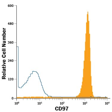
Detection of CD97 in Human Neutrophils by Flow Cytometry.
Human neutrophils were stained with Mouse Anti-Human CD97 APC-conjugated Monoclonal Antibody (Catalog # FAB2529A, filled histogram) or isotype control antibody (Catalog # IC003A, open histogram). View our protocol for Staining Membrane-associated Proteins.Applications for Human CD97 APC-conjugated Antibody
Application
Recommended Usage
Flow Cytometry
10 µL/106 cells
Sample: Human neutrophils
Sample: Human neutrophils
Spectra Viewer
Plan Your Experiments
Use our spectra viewer to interactively plan your experiments, assessing multiplexing options. View the excitation and emission spectra for our fluorescent dye range and other commonly used dyes.
Spectra Viewer
Formulation, Preparation, and Storage
Purification
Protein A or G purified from hybridoma culture supernatant
Formulation
Supplied in a saline solution containing BSA and Sodium Azide.
Shipping
The product is shipped with polar packs. Upon receipt, store it immediately at the temperature recommended below.
Stability & Storage
Store the unopened product at 2 - 8° C. Do not use past expiration date. Protect from light.
Background: CD97
CD97 is a 95‑100 kDa member of a protein group known as the LNB-TM7 protein family that evolved from genes of the secretin receptor superfamily (1‑3). Molecules in this family are unique hybrid structures consisting of EGF-like modules coupled to class B G-protein 7-transmembrane (TM) domains by a glycosylated (mucin) stalk. Human CD97 is synthesized as an 835 amino acid (aa) precursor that contains a 20 aa signal sequence, a 532 aa extracellular domain (ECD), a 238 aa “transmembrane” region that includes seven TM segments, and a 45 aa cytoplasmic tail (4). Within the 532 aa ECD, the first 236 aa contains five EGF-like domains, the C-terminal four of which bind calcium, and a juxtamembrane 296 aa RGD-containing mucin stalk (4, 5). The stalk is both glycosylated and proteolytically cleaved (at aa 530) to create a noncovalently linked 65‑70 kDa glycosylated extracellular alpha-subunit and a 28 kDa 7-TM membrane-bound beta-subunit (4). There are two known alternate splice forms in human. Isoform # 1 contains four EGF-like domains (domain # 1, 2, 3 and 5), while isoform # 2 contains three EGF-like domains (domain # 1, 2 and 5) (1, 4, 6). The ECD in isoform 1 is 60 kDa in size, while the ECD in isoform 2 is 55 kDa in size (native molecular weight). The five EGF-like domain region in human is 55% aa identical to that in mouse. Cells known to express CD97 include monocytes, macrophages, T cells, select B cells, dendritic cells and, potentially, vascular and visceral smooth muscle cells (1, 7). There are at least two ligands for CD97. One is chrondroitin sulfate that binds only to the full-length (five domain) form of CD97. Binding is dependent on the presence of EGF-like domain # 4 (3). The second ligand for CD97 is CD55, a GPI-linked cell surface molecule with short consensus repeats that regulates complement activation on cell surfaces (1, 5, 7). CD97 EGF-like domains # 1 and 2 bind CD55 while domain # 5 stabilizes the CD97 molecule. The shortest CD97 isoform shows the strongest binding to CD55 (7, 8).
References
- McKnight, A.J. and S. Gordon (1998) J. Leukoc. Biol. 63:271.
- Stacey, M. et al. (2000) Trends Biochem. Sci. 25:284.
- Stacey, M. et al. (2003) Blood 102:2916.
- Gray, J.X. et al. (1996) J. Immunol. 157:5438.
- Lin, H-H. et al. (2001) J. Biol. Chem. 276:24160.
- Hamann, J. et al. (1995) J. Immunol. 155:1942.
- Jaspars, L.H. et al. (2001) Tissue Antigens 57:325.
- Hamann, J. et al. (1996) J. Exp. Med. 184:1185.
Alternate Names
CD97
Gene Symbol
ADGRE5
UniProt
Additional CD97 Products
Product Documents for Human CD97 APC-conjugated Antibody
Product Specific Notices for Human CD97 APC-conjugated Antibody
For research use only
Loading...
Loading...
Loading...
Loading...
Loading...