
Human Caspase-9 Antibody
R&D Systems, part of Bio-Techne | Catalog # AF8301

Scientific images may be resized, cropped, or adjusted for brightness or contrast for presentation purposes. These updates are limited to presentation and do not affect the underlying scientific interpretation of the data. Where available, additional source data may be provided upon request.
Conjugate
Catalog #
Key Product Details
Species Reactivity
Validated:
Human
Cited:
Human, Rat
Applications
Validated:
Western Blot
Cited:
Western Blot
Label
Unconjugated
Antibody Source
Polyclonal Goat IgG
Product Specifications
Immunogen
E. coli-derived recombinant human Caspase-9
Val139-Asp330
Accession # P55211
Val139-Asp330
Accession # P55211
Specificity
Detects human Caspase-9 precursor and the pro + large subunit (LSU) that migrates as a 37 kDa band on SDS-PAGE generated by cleavage at Asp330. Does not detect the pro + LSU that migrates as a 34 kDa band on SDS-PAGE generated by cleavage at Asp315. Therefore, the major epitope detected occurs between Asp315 and Asp330.
Clonality
Polyclonal
Host
Goat
Isotype
IgG
Scientific Data Images for Human Caspase-9 Antibody
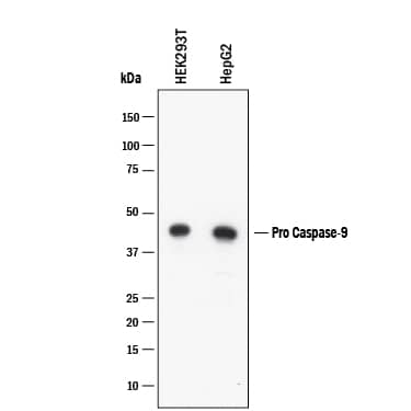
Detection of Human Caspase-9 by Western Blot.
Western blot shows lysates of HEK293T human embryonic kidney cell line and HepG2 human hepatocellular carcinoma cell line. PVDF membrane was probed with 1 µg/mL of Goat Anti-Human Caspase-9 Antigen Affinity-purified Polyclonal Antibody (Catalog # AF8301) followed by HRP-conjugated Anti-Goat IgG Secondary Antibody (Catalog # HAF017). A specific band was detected for Caspase-9 at approximately 46 kDa (as indicated). This experiment was conducted under reducing conditions and using Western Blot Buffer Group 2.
Detection of Human Caspase-9 by Western Blot.
Western blot shows lysates of Jurkat human acute T cell leukemia cell line untreated (-) or treated (+) with 1 ug/ml Staurosporine (STS) for 2 hours. PVDF membrane was probed with 1 µg/mL of Goat Anti-Human Caspase-9 Antigen Affinity-purified Polyclonal Antibody (Catalog # AF8301) followed by HRP-conjugated Anti-Goat IgG Secondary Antibody (Catalog # HAF017). Specific bands were detected for Caspase-9 at approximately 46, 37, 35 kDa (as indicated). This experiment was conducted under reducing conditions and using Western Blot Buffer Group 2.
Detection of Caspase-9 by Western Blot
Effect of STZ injection and MitoTEMPOL on carbonyl and caspase protein levels. (A) Protein carbonylation was increased in the diabetic group compared with the other groups. (B) Representative protein immunoblot of caspase 3, cleaved caspase 3, caspase 9, and cleaved caspase 9 protein. (C) Cleaved caspase 3 protein level was significantly reduced in the diabetic + TEMPOL group compared to the diabetic group only. (D) Cleaved caspase 9 protein level was significantly reduced in the diabetic + TEMPOL group compared to the diabetic group only. Image collected and cropped by CiteAb from the following open publication (https://pubmed.ncbi.nlm.nih.gov/35888150), licensed under a CC-BY license. Not internally tested by R&D Systems.Applications for Human Caspase-9 Antibody
Application
Recommended Usage
Western Blot
1 µg/mL
Sample: Staurosporine-treated Jurkat human acute T cell leukemia cell line, HEK293T human embryonic kidney cell line, HepG2 human hepatocellular carcinoma cell line
Sample: Staurosporine-treated Jurkat human acute T cell leukemia cell line, HEK293T human embryonic kidney cell line, HepG2 human hepatocellular carcinoma cell line
Formulation, Preparation, and Storage
Purification
Antigen Affinity-purified
Reconstitution
Reconstitute at 0.2 mg/mL in sterile PBS. For liquid material, refer to CoA for concentration.
Formulation
Lyophilized from a 0.2 μm filtered solution in PBS with Trehalose. *Small pack size (SP) is supplied either lyophilized or as a 0.2 µm filtered solution in PBS.
Shipping
Lyophilized product is shipped at ambient temperature. Liquid small pack size (-SP) is shipped with polar packs. Upon receipt, store immediately at the temperature recommended below.
Stability & Storage
Use a manual defrost freezer and avoid repeated freeze-thaw cycles.
- 12 months from date of receipt, -20 to -70 °C as supplied.
- 1 month, 2 to 8 °C under sterile conditions after reconstitution.
- 6 months, -20 to -70 °C under sterile conditions after reconstitution.
Background: Caspase-9
Caspase-9 (Cysteine-aspartic acid protease 9/Casp-9; also APAF-3, Mch6 and ICE-LAP6) is a 35-37 kDa member of the peptidase C14A family of enzymes. Casp-9 is an initiator caspase that is part of the intrinsic apoptosis pathway. It is widely expressed and is particularly important during development. Human proCaspase-9 is a 47-48 kDa, 416 amino acid (aa) protein and it contains one CARD region (aa 1-92) and catalytic residues at His237 and Cys287. Following mitochondrial disruption, cytochrome c is released from mitochrondria. Cytochrome c acts on APAF-1, which induces procaspase-9 dimerization. The act of dimerization activates proCasp-9, leading to either the activation of Casp-3, or the autocleavage of proCasp-9, generating a 35 kDa subunit (aa 1-315) and a 12 kDa subunit. Activated Casp-3 will also act on proCasp-9, generating a 37 kDa subunit (aa 1-330) and a 10 kDa subunit (aa 331-416). These subunits associate to form an active heterotetramer. Casp-9 has an alternative start site at Met84 and a deletion of aa 140-289 that generates a dominant negative, 31 kDa isoform. Over aa 1-134, human Casp-9 shares 81% aa identity with mouse Casp-9; over aa 139-330, human Casp-9 shares 73% aa identity with mouse Casp-9.
Additional Caspase-9 Products
Product Documents for Human Caspase-9 Antibody
Product Specific Notices for Human Caspase-9 Antibody
For research use only
Loading...
Loading...
Loading...
Loading...
Loading...