
CD8 Antibody [CoraFluor™ 1]
Novus Biologicals, part of Bio-Techne | Catalog # AF2598CL1

Scientific images may be resized, cropped, or adjusted for brightness or contrast for presentation purposes. These updates are limited to presentation and do not affect the underlying scientific interpretation of the data. Where available, additional source data may be provided upon request.
Conjugate
Catalog #
Key Product Details
Species Reactivity
Feline
Applications
Western Blot, Immunocytochemistry
Label
CoraFluor 1
Antibody Source
Polyclonal Goat IgG
Concentration
Please see the vial label for concentration. If unlisted please contact technical services.
Product Specifications
Immunogen
Mouse myeloma cell line NS0-derived recombinant feline CD8 alpha
Ala22-Tyr188
Accession # P41688
Ala22-Tyr188
Accession # P41688
Specificity
Detects feline CD8 alpha in direct ELISAs and Western blots. In direct ELISAs and Western blots, approximately 25% cross-reactivity with recombinant mouse CD8 alpha is observed, approximately 5% cross-reactivity with recombinant canine CD8 alpha is observed, and less than 1% cross-reactivity with recombinant human CD8 alpha is observed.
Clonality
Polyclonal
Host
Goat
Isotype
IgG
Description
This conjugate is made on demand and is not held in inventory. Each order represents a unique lot. All conjugates are generated using validated conjugation protocols and meet strict degree of labeling (F/P) specifications; additional information is available upon request. The antibody concentration is provided on the individual product label.
For specialized conjugation needs, like large quantities, specific concentrations, and defined F/P ratios, bulk and custom antibody conjugation services are available.
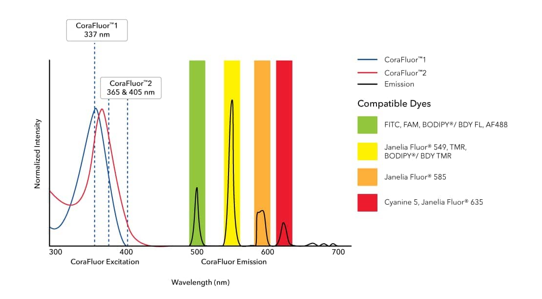
CoraFluor(TM) 1 is a high performance terbium-based TR-FRET (Time-Resolved Fluorescence Resonance Energy Transfer) or TRF (Time-Resolved Fluorescence) donor for high throughput assay development. CoraFluor(TM) 1 absorbs UV light at approximately 340 nm, and emits at approximately 490 nm, 545 nm, 585 nm and 620 nm. It is compatible with common acceptor dyes that absorb at the emission wavelengths of CoraFluor(TM) 1. CoraFluor(TM) 1 can be used for the development of robust and scalable TR-FRET binding assays such as target engagement, ternary complex, protein-protein interaction and protein quantification assays.
CoraFluor(TM) 1, amine reactive
CoraFluor(TM) 1, thiol reactive
For more information, please see our CoraFluor(TM) TR-FRET technology flyer.
Scientific Data Images for CD8 Antibody [CoraFluor™ 1]
Product Feature: CoraFluor Probes for TR-FRET
CoraFluor™ 1, amine reactive (Catalog:7920) and CoraFluor™ 2, amine reactive (Catalog # 7950) are terbium-based probes that have been developed for use as TR-FRET donors. They emit wavelengths compatible with commonly used fluorescent acceptor dyes such as BODIPY® (or BDY) and Janelia Fluor® dyes, FITC (Catalog # 5440), TMR and Cyanine 5 (Catalog # 5436). CoraFluor™ fluorescence is brighter and more stable in biological media than existing TR-FRET donors, leading to enhanced sensitivity and improved data generation. CoraFluor™ 1 exhibits excitation upon exposure to a 337 nm UV laser.Applications for CD8 Antibody [CoraFluor™ 1]
Application
Recommended Usage
Immunocytochemistry
Optimal dilutions of this antibody should be experimentally determined.
Western Blot
Optimal dilutions of this antibody should be experimentally determined.
Application Notes
Recommended applications are based on validated applications from the unconjugated base product AF2598. This conjugated antibody is not kept in inventory and is made to order using validated conjugation protocols.
Formulation, Preparation, and Storage
Purification
Antigen Affinity-purified
Formulation
PBS
Preservative
No Preservative
Concentration
Please see the vial label for concentration. If unlisted please contact technical services.
Shipping
The product is shipped with polar packs. Upon receipt, store it immediately at the temperature recommended below.
Stability & Storage
Store at 4C in the dark. Do not freeze.
Background: CD8
Given its role in the immune system, CD8-deficiency in T-cells is a hallmark of many diseases and pathologies (8-10). Specifically, CD8+ T-cell deficiency is prevalent in chronic autoimmune diseases including multiple sclerosis, rheumatoid arthritis, ulcerative colitis, Crohn's disease, type 1 diabetes mellitus, and Graves' disease (8). Furthermore, cancers or chronic infection can lead to CD8 T-cell exhaustion as the continual antigen presentation and inflammatory signals eventually cause the CD8+ T-cells to lose functionality (9, 10). However, animal models and clinical studies have suggested that T-cells are capable of being reinvigorated using inhibitory receptor blockade resulting in better disease outcomes and these exhausted T-cells may be a potential therapeutic target (9, 10).
Alternative names for CD8 includes CD antigen: CD8a, CD8 antigen, alpha polypeptide (p32), CD8a molecule, CD8A, Leu2 T-lymphocyte antigen, LEU2, MAL, OKT8 T-cell antigen, p32, T cell co-receptor, T8 T-cell antigen, T-cell antigen Leu2, T-cell surface glycoprotein CD8 alpha chain, and T-lymphocyte differentiation antigen T8/Leu-2.
References
1. Littman D. R. (1987). The structure of the CD4 and CD8 genes. Annual review of immunology. https://doi.org/10.1146/annurev.iy.05.040187.003021
2. Naeim F. (2008). Chapter 2- Principles of Immunophenotyping. Hematopathology. https://doi.org/10.1016/B978-0-12-370607-2.00002-8.
3. Gao, G. F., & Jakobsen, B. K. (2000). Molecular interactions of coreceptor CD8 and MHC class I: the molecular basis for functional coordination with the T-cell receptor. Immunology today. https://doi.org/10.1016/s0167-5699(00)01750-3
4. UniProt (P01732)
5. UniProt (P01731)
6. Kappes D. J. (2007). CD4 and CD8: hogging all the Lck. Immunity. https://doi.org/10.1016/j.immuni.2007.11.002
7. Gangadharan, D., & Cheroutre, H. (2004). The CD8 isoform CD8alphaalpha is not a functional homologue of the TCR co-receptor CD8alphabeta. Current opinion in immunology. https://doi.org/10.1016/j.coi.2004.03.015
8. Pender M. P. (2012). CD8+ T-Cell Deficiency, Epstein-Barr Virus Infection, Vitamin D Deficiency, and Steps to Autoimmunity: A Unifying Hypothesis. Autoimmune diseases. https://doi.org/10.1155/2012/189096
9. Kurachi M. (2019). CD8+ T cell exhaustion. Seminars in immunopathology. https://doi.org/10.1007/s00281-019-00744-5
10. Hashimoto, M., Kamphorst, A. O., Im, S. J., Kissick, H. T., Pillai, R. N., Ramalingam, S. S., Araki, K., & Ahmed, R. (2018). CD8 T Cell Exhaustion in Chronic Infection and Cancer: Opportunities for Interventions. Annual review of medicine. https://doi.org/10.1146/annurev-med-012017-043208
Alternate Names
CD8, CD8A
Gene Symbol
CD8A
Additional CD8 Products
Product Documents for CD8 Antibody [CoraFluor™ 1]
Product Specific Notices for CD8 Antibody [CoraFluor™ 1]
Sold for research purposes only under agreement from Massachusetts General Hospital, US Patent No. 12,134,721. A license is required for development of new commercial assay platforms (e.g., microarrays, microfluidics), diagnostic use or other unlicensed uses. Please contact Technical Support for further details: techsupport@bio-techne.com.
This product is for research use only and is not approved for use in humans or in clinical diagnosis. Primary Antibodies are guaranteed for 1 year from date of receipt.
Loading...
Loading...
Loading...
Loading...