Peripheral Node Addressin Antibody (MECA-79R) - BSA Free
Novus Biologicals, part of Bio-Techne | Catalog # NBP2-78792


Conjugate
Catalog #
Forumulation
Catalog #
Key Product Details
Species Reactivity
Validated:
Human, Mouse
Cited:
Human, Mouse
Applications
Validated:
Immunocytochemistry/ Immunofluorescence, Immunohistochemistry, Immunohistochemistry-Frozen, Immunohistochemistry-Paraffin, In vivo assay, Multiplex Immunofluorescence, Western Blot
Cited:
IF/IHC, Immunocytochemistry/ Immunofluorescence, Immunohistochemistry, Immunohistochemistry-Frozen, Immunohistochemistry-Paraffin, In vivo assay, Multiplex Immunoassay
Label
Unconjugated
Antibody Source
Monoclonal Rat IgG1 Clone # MECA-79R
Format
BSA Free
Concentration
1.0 mg/ml
Product Specifications
Immunogen
Mouse lymph node stromal cells
Clonality
Monoclonal
Host
Rat
Isotype
IgG1
Description
Clone MECA-79R (rat IgG1) is a recombinant version of the original clone MECA-79 (rat IgM).
Scientific Data Images for Peripheral Node Addressin Antibody (MECA-79R) - BSA Free
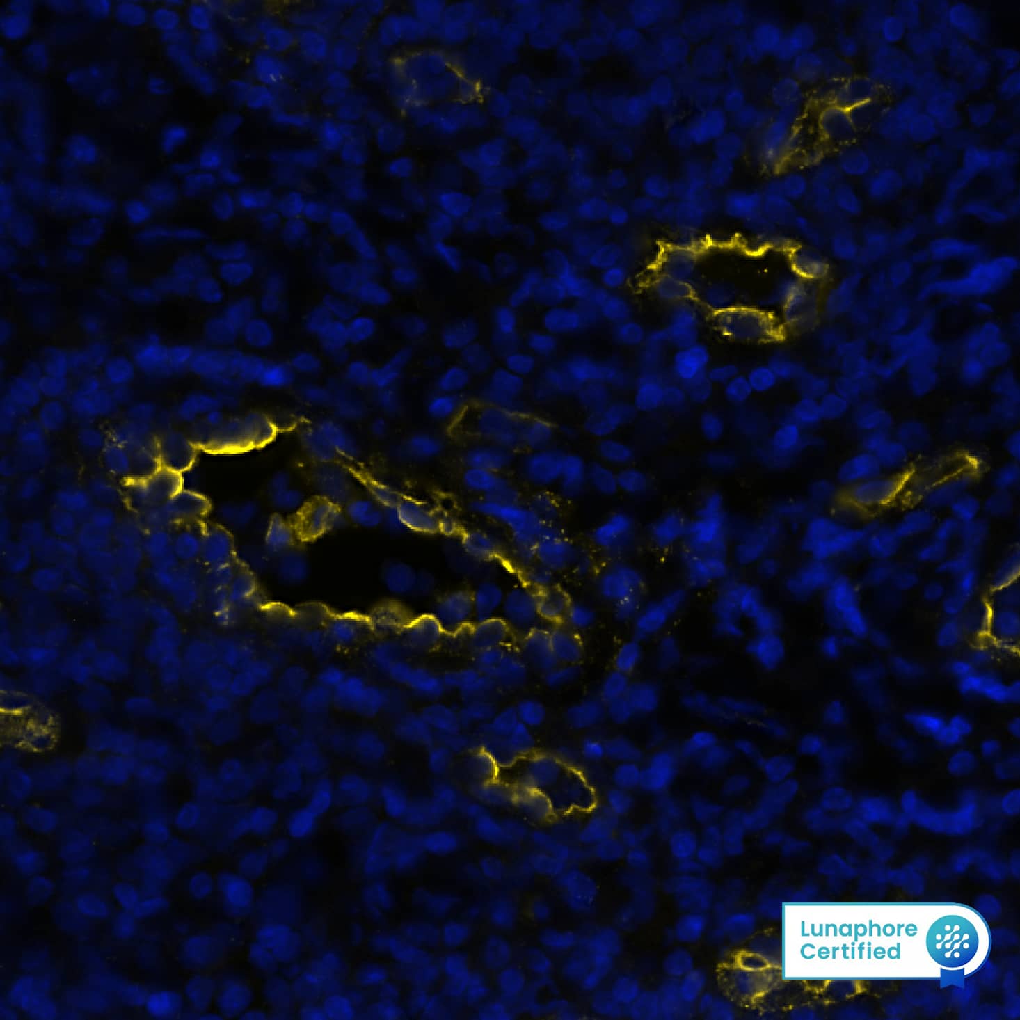
Detection of Peripheral Node Addressin in Human Tonsil via seqIF™ staining on COMET™
Peripheral Node Addressin was detected in immersion fixed paraffin-embedded sections of human Tonsil using Rat Anti-Human Peripheral Node Addressin (MECA-79R), Monoclonal Antibody (Catalog #NBP2-78792) at 1:50 dilution at 37° Celsius for 4 minutes. Before incubation with the primary antibody, tissue underwent an all-in-one dewaxing and antigen retrieval preprocessing using PreTreatment Module (PT Module) and Dewax and HIER Buffer H (pH 9; Epredia Catalog # TA-999-DHBH). Tissue was stained using the Alexa Fluor™ 647 Goat anti-Rat IgG Secondary Antibody at 1:200 at 37 ° Celsius for 2 minutes. (Yellow; Lunaphore Catalog # DR647RT) and counterstained with DAPI (blue; Lunaphore Catalog # DR100). Specific staining was localized to endothelial cells. Protocol available in COMET™ Panel Builder.Western Blot: Peripheral Node Addressin Antibody (MECA-79R)BSA Free [NBP2-78792]
Western Blot: Peripheral Node Addressin Antibody (MECA-79R) [NBP2-78792] - Total protein from human Tonsil, Lymph node, Spleen and mouse Spleen was separated on a 7.5% gel by SDS-PAGE, transferred to PVDF membrane and blocked in 5% non-fat milk in TBST. The membrane was probed with 2.0 ug/ml anti-PNAd in blocking buffer and detected with an anti-rat HRP secondary antibody using West Pico PLUS chemiluminescence detection reagent.Immunohistochemistry-Paraffin: Peripheral Node Addressin Antibody (MECA-79R) - BSA Free [NBP2-78792]
Immunohistochemistry-Paraffin: Peripheral Node Addressin Antibody (MECA-79R) [NBP2-78792] - Analysis of FFPE mouse adipose tissue section (with lymph node areas) using Peripheral Node Addressin antibody (clone MECA-79) at 1:100. The staining was developed with HRP-DAB detection method and the counterstaining was performed using hematoxylin. This Peripheral Node Addressin antibody generated a strong and specific staining of MECA-79 antigen in the the cytoplasm and the membranes of high endothelial venules (HEVs) aka peripheral lymph node addressin (PNAd) in lymph node areas of tested adipose tissue section.Applications for Peripheral Node Addressin Antibody (MECA-79R) - BSA Free
Application
Recommended Usage
Immunocytochemistry/ Immunofluorescence
reported in scientific literature (PMID 31278331)
Immunohistochemistry
1:100-1:500
Immunohistochemistry-Frozen
1:100-1:500
Immunohistochemistry-Paraffin
1:100-1:500
In vivo assay
reported in scientific literature (PMID 30277476)
Multiplex Immunofluorescence
1:50
Western Blot
1:100-1:2000
Application Notes
Additional reported application of in vitro and in vivo blocking of cell adhesion.
Formulation, Preparation, and Storage
Purification
Protein A or G purified
Formulation
PBS
Format
BSA Free
Preservative
0.02% Sodium Azide
Concentration
1.0 mg/ml
Shipping
The product is shipped with polar packs. Upon receipt, store it immediately at the temperature recommended below.
Stability & Storage
Store at -20 °C.
Background: Peripheral Node Addressin
Alternate Names
MECA-79, Peripheral Node Addressin, PNAd
Additional Peripheral Node Addressin Products
Product Documents for Peripheral Node Addressin Antibody (MECA-79R) - BSA Free
Product Specific Notices for Peripheral Node Addressin Antibody (MECA-79R) - BSA Free
This product is for research use only and is not approved for use in humans or in clinical diagnosis. Primary Antibodies are guaranteed for 1 year from date of receipt.
Loading...
Loading...
Loading...
Loading...
![Western Blot: Peripheral Node Addressin Antibody (MECA-79R)BSA Free [NBP2-78792] Western Blot: Peripheral Node Addressin Antibody (MECA-79R)BSA Free [NBP2-78792]](https://resources.bio-techne.com/images/products/Peripheral-Node-Addressin-Antibody-MECA-79R-Western-Blot-NBP2-78792-img0002.jpg)
![Immunohistochemistry-Paraffin: Peripheral Node Addressin Antibody (MECA-79R) - BSA Free [NBP2-78792] Immunohistochemistry-Paraffin: Peripheral Node Addressin Antibody (MECA-79R) - BSA Free [NBP2-78792]](https://resources.bio-techne.com/images/products/Peripheral-Node-Addressin-Antibody-MECA-79R-Immunohistochemistry-Paraffin-NBP2-78792-img0003.jpg)
![Immunohistochemistry-Paraffin: Peripheral Node Addressin Antibody (MECA-79R) - BSA Free [NBP2-78792] Immunohistochemistry-Paraffin: Peripheral Node Addressin Antibody (MECA-79R) - BSA Free [NBP2-78792]](https://resources.bio-techne.com/images/products/Peripheral-Node-Addressin-Antibody-MECA-79R-Immunohistochemistry-Paraffin-NBP2-78792-img0001.jpg)
![Immunohistochemistry: Peripheral Node Addressin Antibody (MECA-79R) - BSA Free [NBP2-78792] Immunohistochemistry: Peripheral Node Addressin Antibody (MECA-79R) - BSA Free [NBP2-78792]](https://resources.bio-techne.com/images/products/Peripheral-Node-Addressin-Antibody-MECA-79R-Immunohistochemistry-NBP2-78792-img0004.jpg)