Human S100A4 Antibody
R&D Systems, part of Bio-Techne | Catalog # MAB4137


Key Product Details
Validated by
Species Reactivity
Validated:
Cited:
Applications
Validated:
Cited:
Label
Antibody Source
Product Specifications
Immunogen
Met1-Ser101
Accession # P26447
Specificity
Clonality
Host
Isotype
Scientific Data Images for Human S100A4 Antibody
Detection of Human and Mouse S100A4 by Western Blot.
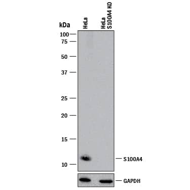
Western blot shows lysates of HeLa human cervical epithelial carcinoma cell line and NIH-3T3 mouse embryonic fibroblast cell line. PVDF membrane was probed with 2 µg/mL of Mouse Anti-Human S100A4 Monoclonal Antibody (Catalog # MAB4137) followed by HRP-conjugated Anti-Mouse IgG Secondary Antibody (Catalog # HAF018). A specific band was detected for S100A4 at approximately 11 kDa (as indicated). This experiment was conducted under reducing conditions and using Immunoblot Buffer Group 1.S100A4 in Human Cervical Cancer Tissue.
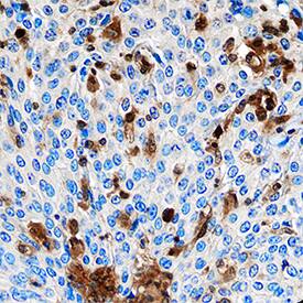
S100A4 was detected in immersion fixed paraffin-embedded sections of human cervical cancer tissue using Mouse Anti-Human S100A4 Monoclonal Antibody (Catalog # MAB4137) at 15 µg/mL overnight at 4 °C. Tissue was stained using the Anti-Mouse HRP-DAB Cell & Tissue Staining Kit (brown; Catalog # CTS002) and counterstained with hematoxylin (blue). Specific staining was localized to cytoplasm in cancer cells. View our protocol for Chromogenic IHC Staining of Paraffin-embedded Tissue Sections.S100A4 in A549 Human Cell Line.
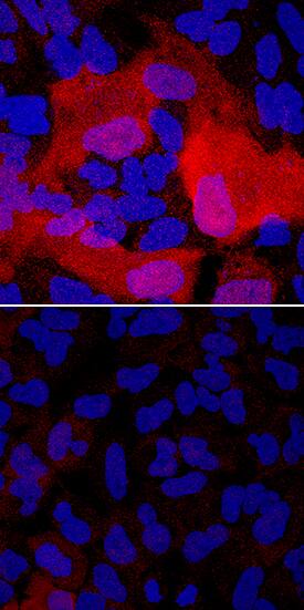
S100A4 was detected in immersion fixed A549 human lung carcinoma cells unstimulated (lower panel) and stimulated with the EMT Inducing Media Supplement (upper panel; Catalog # CCM017) using Mouse Anti-Human S100A4 Monoclonal Antibody (Catalog # MAB4137) at 1 µg/mL for 3 hours at room temperature. Cells were stained using the NorthernLights™ 557-conjugated Anti-Mouse IgG Secondary Antibody (red; Catalog # NL007) and counterstained with DAPI (blue). Specific staining was localized to cytoplasm. View our protocol for Fluorescent ICC Staining of Cells on Coverslips.Applications for Human S100A4 Antibody
Immunocytochemistry
Sample: Immersion fixed A549 human lung carcinoma cells unstimulated and stimulated with the EMT Inducing Media Supplement (Catalog # CCM017)
Immunohistochemistry
Sample: Immersion fixed paraffin-embedded sections of human cervical cancer tissue
Knockout Validated
Western Blot
Sample: HeLa human cervical epithelial carcinoma cell line and NIH‑3T3 mouse embryonic fibroblast cell line
Reviewed Applications
Read 4 reviews rated 4.5 using MAB4137 in the following applications:
Formulation, Preparation, and Storage
Purification
Reconstitution
Formulation
Shipping
Stability & Storage
- 12 months from date of receipt, -20 to -70 °C as supplied.
- 1 month, 2 to 8 °C under sterile conditions after reconstitution.
- 6 months, -20 to -70 °C under sterile conditions after reconstitution.
Background: S100A4
S100A4, also known as Metastasin, Mtsl and Calvasculin, is an 11 kDa member of the S100 (soluble in 100% saturated ammonium sulfate) family of proteins (1-5). The S100 family is further classified as a member of the EF-hand superfamily of Ca++-binding proteins. These participate in both calcium-dependent and
calcium-independent protein-protein interactions. The hallmark of this superfamily is the EF-hand motif that consists of a Ca++-binding site flanked by two alpha-helices (helix E and helix F) that were originally identified in a right-handed model of carp muscle calcium-binding protein (6). Human S100A4 is 101 amino acids (aa) in length (1, 2). It contains two EF hand domains, one between aa 12-47, and a second between aa 50-85. The first domain has a 14 aa cation-binding motif and binds Ca++ with low affinity. The second Ca++-binding motif is 12 aa in length and binds Ca++ with high affinity. S100A4 has no classical signal sequence but is secreted from cells (3, 7). Human S100A4 shares 93% and 95% aa identity with mouse and canine S100A4, respectively. S100A4 reportedly exists as a dimer (8, 9, 10). Extracellular S100A4 is reported to induce MMP production, activate MMPs, promote neurite outgrowth and stimulate cardiomyocyte proliferation (4, 10, 11, 12, 13). Within the cell, dimers are likely the functional unit. Here, they are constitutive homo- or heterodimers (with S100A1) that interact with Ca++, undergo a conformational change, and subsequently bind to cytoplasmic targets. Known targets include p53, myosin heavy chain II, F-actin and liprin beta1 (4, 14). In general, it can be said that S100A4 blocks target phosphorylation and multimerization (4, 7, 14). S100A4 activity has been associated with cell transformation. It seems likely this is either coincidental, or a consequence, rather than a cause of transformation (3).
References
- Engelkamp, D. et al. (1992) Biochemistry 31:10258.
- Tomida, Y. et al. (1992) Biochem. Biophys. Res. Commun. 189:1310.
- Garrett, S.C. et al. (2006) J. Biol. Chem. 281:677.
- Santamaria-Kisiel, L. et al. (2006) Biochem. J. 396:201.
- Donato, R. (2001) Int. J. Biochem. Mol. Biol. 33:637.
- Kretsinger, R.H. and C.E. Nockolds (1973) J. Biol. Chem. 248:3313.
- Helfman, D.M. et al. (2005) Br. J. Cancer 92:1955.
- Burkitt, W.I. et al. (2003) Biochem. Soc. Trans. 31:985.
- Vallaly, K.M. et al. (2002) Biochemistry 41:12670.
- Novitskaya, V. et al. (2000) J. Biol. Chem. 275:41278.
- Stary, M. et al. (2006) Biochem. Biophys. Res. Commun. 343:555.
- Semov, A. et al. (2005) J. Biol. Chem. 280:20833.
- Saleem, M. et al. (2006) Proc. Natl. Acad. Sci. USA 103:14825.
- Kriajevska, M. et al. (2002) J. Biol. Chem. 277:5229.
Long Name
Alternate Names
Gene Symbol
UniProt
Additional S100A4 Products
Product Documents for Human S100A4 Antibody
Product Specific Notices for Human S100A4 Antibody
For research use only


