Human KGF/FGF-7 Antibody
R&D Systems, part of Bio-Techne | Catalog # AF-251-NA


Key Product Details
Validated by
Species Reactivity
Validated:
Cited:
Applications
Validated:
Cited:
Label
Antibody Source
Product Specifications
Immunogen
Cys32-Thr194
Accession # Q6FGV5
Specificity
Clonality
Host
Isotype
Endotoxin Level
Scientific Data Images for Human KGF/FGF-7 Antibody
Cell Proliferation Induced by KGF/FGF‑7 and Neutralization by Human KGF/FGF‑7 Antibody.
Recombinant Human KGF/FGF-7 (Catalog # 251-KG) stimulates proliferation in the 4MBr-5 rhesus monkey epithelial cell line in a dose-dependent manner (orange line). Proliferation elicited by Recombinant Human KGF/FGF-7 (125 ng/mL) is neutralized (green line) by increasing concentrations of Goat Anti-Human KGF/FGF-7 Antigen Affinity-purified Polyclonal Antibody (Catalog # AF-251-NA). The ND50 is typically 6-12 µg/mL.KGF/FGF‑7 in Human Placenta.
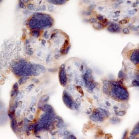
KGF/FGF-7 was detected in immersion fixed paraffin-embedded sections of human placenta (chorionic villi) using Goat Anti-Human KGF/FGF-7 Antigen Affinity-purified Polyclonal Antibody (Catalog # AF-251-NA) at 10 µg/mL overnight at 4 °C. Before incubation with the primary antibody, tissue was subjected to heat-induced epitope retrieval using Antigen Retrieval Reagent-Basic (Catalog # CTS013). Tissue was stained using the Anti-Goat HRP-DAB Cell & Tissue Staining Kit (brown; Catalog # CTS008) and counterstained with hematoxylin (blue). Specific staining was localized to trophoblast cells in chorionic villi. View our protocol for Chromogenic IHC Staining of Paraffin-embedded Tissue Sections.Detection of Porcine KGF/FGF-7 by Immunohistochemistry-Paraffin
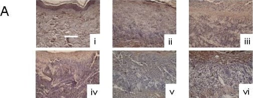
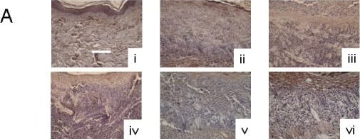
Immunohistochemical detection of KGF-2 in biopsies of DNCB-induced rashes treated with various nanocrystalline silver-derived solutions. (A) Representative images are shown for immunohistochemical detection of KGF-2 after 24 h treatment of negative controls with distilled water (i), and DNCB-induced porcine contact dermatitis rashes with distilled water (positive controls) (ii), or nanocrystalline silver-derived solutions generated at starting pHs of 4 (iii), 5.6 (iv), 7 (v), or 9 (vi). The scale bar in A represents 100 μm. Staining for KGF-2 appears brown, while the cell nuclei are counterstained purple using haematoxylin. Immunohistochemical staining scores for KGF-2 are shown after 24 h (B) and 72 h (C) of treatment as described above. Statistical analyses, which were performed using one-way ANOVAs with Tukey-Kramer Multiple Comparisons post tests, are shown in Table 4. Error bars represent standard deviations. Image collected and cropped by CiteAb from the following open publication (https://pubmed.ncbi.nlm.nih.gov/20170497), licensed under a CC-BY license. Not internally tested by R&D Systems.Applications for Human KGF/FGF-7 Antibody
Immunohistochemistry
Sample: Immersion fixed paraffin-embedded sections of human placenta
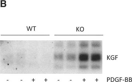
Western Blot
Sample: Recombinant Human KGF/FGF-7 (Catalog # 251-KG)
Neutralization
Reviewed Applications
Read 2 reviews rated 4 using AF-251-NA in the following applications:
Formulation, Preparation, and Storage
Purification
Reconstitution
Formulation
Shipping
Stability & Storage
- 12 months from date of receipt, -20 to -70 °C as supplied.
- 1 month, 2 to 8 °C under sterile conditions after reconstitution.
- 6 months, -20 to -70 °C under sterile conditions after reconstitution.
Background: KGF/FGF-7
KGF (keratinocyte growth factor), also known as FGF-7 (fibroblast growth factor-7), is one of 22 known members of the mouse FGF family of secreted proteins that plays a key role in development, morphogenesis, angiogenesis, wound healing, and tumorigenesis (1‑4). KGF expression is restricted to cells of mesenchymal origin. When secreted, it acts as a paracrine growth factor for nearby epithelial cells (1). KGF speeds wound healing by being dramatically upregulated in response to damage to skin or internal structures that results in high local concentrations of inflammatory mediators such as IL-1 and TNF-alpha. (2, 5). KGF promotes cell migration and invasion, and mediates melanocyte transfer to keratinocytes upon UVB radiation (6, 7). It has been used ectopically to avoid chemotherapy-induced oral mucositis in patients with hematological malignancies (1). Deletion of KGF affects kidney development, producing abnormally small ureteric buds and fewer nephrons (8). It also impedes hair follicle differentiation (9). The 194 amino acid (aa) KGF precursor contains a 31 aa signal sequence and, like all other FGFs, an ~120 aa beta-trefoil scaffold that includes receptor- and heparin-binding sites. KGF signals only through the IIIb splice form of the tyrosine kinase receptor, FGF R2 (FGF R2-IIIb/KGF R) (10). Receptor dimerization requires an octameric or larger heparin or heparin sulfate proteoglycan (11). FGF-10, also called KGF2, shares 51% aa identity and similar function to KGF, but shows more limited expression than KGF and uses an additional receptor, FGF R2-IIIc (12). Following receptor engagement, KGF is typically degraded, while FGF-10 is recycled (12). Mature human KGF, which is active across species, shares 98% aa sequence identity with bovine, equine, ovine and canine, 96% with mouse and porcine, and 92% with rat KGF, respectively.
References
- Finch, P.W. and J.S. Rubin (2006) J. Natl. Cancer Inst. 98:812.
- Werner, S. et al. (2007) J. Invest. Dermatol. 127:998.
- Werner, S. (1998) Cytokine Growth Factor Rev. 9:153.
- Mason, I.J. et al. (1994) Mech. Dev. 45:15.
- Geer, D.J. et al. (2005) Am. J. Pathol. 167:1575.
- Niu, J. et al. (2007) J. Biol. Chem. 282:6001.
- Cardinali, G. et al. (2005) J. Invest. Dermatol. 125:1190.
- Qiao, J. et al. (1999) Development 126:547.
- Guo, L. et al. (1996) Genes Dev. 10:165.
- de Georgi, V. et al. (2007) Dermatol. Clin. 25:477.
- Hsu, Y-R. et al. (1999) Biochemistry 38:2523.
- Belleudi, F. et al. (2007) Traffic 8:1854.
Long Name
Alternate Names
Gene Symbol
UniProt
Additional KGF/FGF-7 Products
Product Documents for Human KGF/FGF-7 Antibody
Product Specific Notices for Human KGF/FGF-7 Antibody
For research use only


