Mouse CD4+ T Cell Enrichment Column, Small, Pack of 4
Key Product Details
Order Information
Shipping Information
$35 to United States
Products in stock will be shipped in 1-3 business days unless specified.
Assay Procedure
Refer to the product datasheet for complete product details.
Briefly, mouse CD4+ T cells can be isolated using the following procedure:
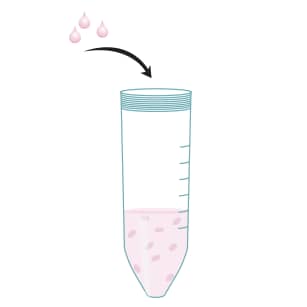
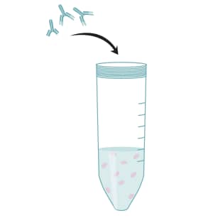
- Incubate a single-cell suspension of leukocytes with the Monoclonal Antibody Cocktail
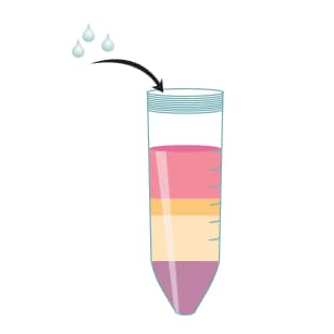
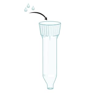
- Apply antibody-treated cell suspension to the CD4+ T Cell Subset Column
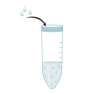
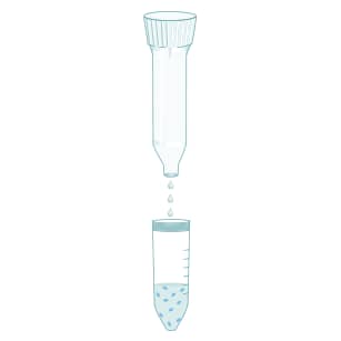
- Elute CD4+ T cells with 1X Column Buffer
Kit Contents
The Mouse CD4+ T Cell Enrichment Column Kit, Small (Catalog #MCD4C-1000) contains the following components to allow for the isolation of mouse CD4+ T cells.
- Mouse CD4+ T Cell Subset Columns
- Monoclonal Antibody Cocktail
- Column Buffer Concentrate (10X)
Other Supplies Required
Technical notes
- In order to best determine column performance, we advise that users retain a small portion of the starting cell population. Following cell selection with the column, it is then possible to perform immunophenotyping analysis on both starting and eluted cells. This information, when combined with the actual number of cells loaded and recovered, can then be used to calculate the percentage recovery of the target cell population.
- Care should be taken during cell preparation to reduce the number of platelets before loading the cells onto the column. Platelet contamination can be reduced by performing a final wash centrifugation of the cells at a reduced speed (150 - 200 x g).
- Some of the salts in the 10X column buffer solution may precipitate after storage at 4 °C. Should this be the case, do not carry out the 1:10 buffer dilution until all salts are in solution. This may be achieved by warming the 10X column buffer bottle in a 37 °C water bath for 5 - 10 minutes. Once there is no longer evidence of precipitates, the 10X column buffer may be diluted 1:10 to prepare the 1X column buffer for column processing.
Helpful Hints in Running T Cell Enrichment Columns
- Remove as many clumps as possible from the cell suspension before loading cells onto the column. Although the column is designed to filter out larger clumps of cells, too many clumps on the filter will reduce the column flow rate and cell recovery. In addition, leaving a large number of cells in a small volume of buffer for more than 30 minutes may promote cell clumping.
- If cells do not move into column after 15 minutes, the filter may have become clogged. If this occurs, move the white filter at the top of the column to the side with a sterile pipette. The cells should migrate into the column more easily.
- The column is designed so that the white filter at the top of the column bed will stop buffer flow and prevent the column from drying out. However leaving the open column exposed to air for more than 1 hour may cause the column bed to dry out and the CD4+ T cell enrichment to be compromised.
- Cell recovery after column processing is largely dependent on the total number of cells loaded. Optimal column performance is achieved by loading approximately 75 x106 cells. Loading fewer cells will negatively affect total cell recovery.
- If buffer does not drip out of column after initial removal of the bottom cap, try tapping the side of the column to remove any air locks that may be preventing the flow of buffer.
- The white filter at the top of the column bed may be found floating as a result of being dislodged during shipping. The column can still be used for cell processing after pushing the white filter onto the column bed with the back end of a sterile 2 mL pipette. The white filter should be positioned close to the column bed.
Procedure Overview
R&D Systems Protocol for Mouse CD4+ T Cell Enrichment Column, Small
Prepare a single cell suspension of mononuclear cells.
Wash the cells with excess sterile PBS.

Remove the red blood cells if necessary (e.g., using R&D Systems Mouse Erythrocyte Lysing Kit Catalog # WL2000)
Resuspend the cells in a small volume of 1X Column Wash Buffer.

Perform a cell count.
Adjust the cell concentration to 1-2 x 10⁸ cells/mL with 1X Column Wash Buffer.

Mix 2 x 10⁸ suspended cells with the contents of one vial of Monoclonal Antibody Cocktail.
Incubate for 15 minutes at room temperature.

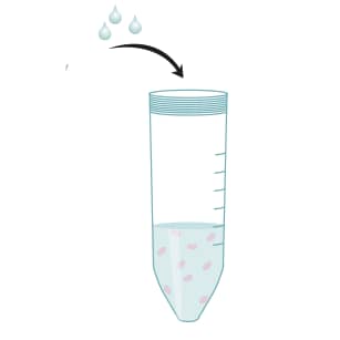
Wash cells twice with 10 mL 1X Column Wash Buffer.
Resuspend cells in 2 mL of 1X Column Wash Buffer.

Load cell suspension onto the prepared column.
Incubate for 10 minutes at room temperature.

Elute CD4+ T cells with 10 mL of 1X Column Wash Buffer.
Collect the cells in a sterile 50 mL centrifuge tube.

Pellet the cells using centrifugation.
Resuspend CD4+ T cells in the appropriate buffer or culture medium for downstream applications.

Citations for Mouse CD4+ T Cell Enrichment Column, Small, Pack of 4 (21)
Citations are publications that use Bio-Techne products. Selected citations for Mouse CD4+ T Cell Enrichment Column, Small, Pack of 4 include:
-
Species: Human
Sample Types: Whole Cells
A Romano et al. (2020), High-density neutrophils in MGUS and multiple myeloma are dysfunctional and immune-suppressive due to increased STAT3 downstream signaling Sci Rep, 10(1):1983.
PMID: 32029833 -
Species: Mouse
Sample Types: Whole Cells
A Taylor et al. (2020), Glycogen synthase kinase 3 (GSK-3) controls T-cell motility and interactions with antigen presenting cells BMC Res Notes, 13(1):163.
PMID: 32188506 -
Species: Mouse
Sample Types: Whole Cells
F Muhammad et al. (2019), PD-1+ melanocortin receptor dependent-Treg cells prevent autoimmune disease Sci Rep, 9(1):16941.
PMID: 31729418 -
Species: Mouse
Sample Types: Whole Cells
D Erturk-Has et al. (2019), Symbionts exploit complex signaling to educate the immune system Proc. Natl. Acad. Sci. U.S.A., 0(0).
PMID: 31811024 -
Species: Mouse
Sample Types: Whole Cells
K Shimano et al. (2019), Amiselimod (MT-1303), a novel sphingosine 1-phosphate receptor-1 functional antagonist, inhibits progress of chronic colitis induced by transfer of CD4+CD45RBhigh�T cells PLoS ONE, 14(12):e0226154.
PMID: 31805144 -
Species: Mouse
Sample Types: Whole Cells
Y Zhao et al. (2019), High Thymic Output of Effector CD4+ Cells May Lead to a Treg?:?T Effector Imbalance in the Periphery in NOD Mice J Immunol Res, 2019(0):8785263.
PMID: 31281853 -
Species: Mouse
Sample Types: Whole Cells
Q Chen et al. (2019), ?-Galactosylceramide treatment before allergen sensitization promotes iNKT cell-mediated induction of Treg cells, preventing Th2 cell responses in murine asthma J. Biol. Chem., 0(0).
PMID: 30745361 -
Species: Mouse
Sample Types: Whole Cells
Barbara L F Kaplan (2016), Cannabidiol (CBD) induces functional Tregs in response to low-level T cell activation Cell. Immunol., 0(0).
PMID: 27865421 -
Species: Mouse
Sample Types: Whole Cells
Jessica Harakal (2016), Regulatory T Cells Control Th2-Dominant Murine Autoimmune Gastritis J Immunol, 197(1):27-41.
PMID: 27259856 -
Species: Mouse
Sample Types: Whole Cells
(2016), Defect density in multiwalled carbon nanotubes influences ovalbumin adsorption and promotes macrophage activation and CD4(+) T-cell proliferation Int J Nanomedicine, 11(0):4357-71.
PMID: 27621627 -
Species: Mouse
Sample Types: Whole Cells
(2016), Digoxin Inhibits Induction of Experimental Autoimmune Uveitis in Mice, but Causes Severe Retinal Degeneration Invest. Ophthalmol. Vis. Sci., 57(3):1441-7.
PMID: 27028065 -
Species: Mouse
Sample Types: Whole Cells
Kant C et al. (2015), Both rejection and tolerance of allografts can occur in the absence of secondary lymphoid tissues. J Immunol, 194(3):1364-71.
PMID: 25535285 -
Species: Mouse
Sample Types: Whole Cells
Kim H et al. (2013), Dynamic motile T cells highly respond to the T cell stimulation via PI3K-Akt and NF-kappaB pathways. PLoS ONE, 8(3):e59793.
PMID: 23555783 -
Species: Mouse
Sample Types: Whole Cells
Surls J et al. (2012), Increased membrane cholesterol in lymphocytes diverts T-cells toward an inflammatory response. PLoS ONE, 7(6):e38733.
PMID: 22723880 -
Species: Mouse
Sample Types: Whole Cells
Tan C et al. (2010), Antigen-specific Th9 cells exhibit uniqueness in their kinetics of cytokine production and short retention at the inflammatory site. J. Immunol., 185(11):6795-801.
PMID: 20971929 -
Species: Mouse
Sample Types: Whole Cells
Wallace KL et al. (2009), NKT cells mediate pulmonary inflammation and dysfunction in murine sickle cell disease through production of IFN-gamma and CXCR3 chemokines. Blood, 114(3):667-76.
PMID: 19433855 -
Species: Mouse
Sample Types: Whole Cells
Fujiwara M et al. (2007), T-bet inhibits both TH2 cell-mediated eosinophil recruitment and TH17 cell-mediated neutrophil recruitment into the airways. J. Allergy Clin. Immunol., 119(3):662-70.
PMID: 17336616 -
Species: Mouse
Sample Types: Whole Cells
Higgins SC et al. (2006), TLR4 mediates vaccine-induced protective cellular immunity to Bordetella pertussis: role of IL-17-producing T cells. J. Immunol., 177(11):7980-9.
PMID: 17114471 -
Species: Mouse
Sample Types: Whole Cells
De Rosa V et al. (2006), Leptin neutralization interferes with pathogenic T cell autoreactivity in autoimmune encephalomyelitis. J. Clin. Invest., 116(2):447-55.
PMID: 16410832 -
Species: Mouse
Sample Types: Whole Cells
Chen et al. (2003), Conversion of peripheral CD4+CD25- naive T cells to CD4+CD25+ regulatory T cells by TGF-beta induction of transcription factor Foxp3. J Exp Med, 198(12):1875-86.
PMID: 14676299 -
Species: Mouse
Sample Types: Whole Cells
Thornton et al. (1998), CD4+CD25+ immunoregulatory T cells suppress polyclonal T cell activation in vitro by inhibiting interleukin 2 production. J Exp Med, 188(2):287-96.
PMID: 9670041
There are no citations that match your criteria.
Customer Reviews (1)
Customer Images
-
Verified Customer | Posted 10/13/2017Have been using this kit in our laboratory for over 6 years in order to isolate CD4+ T cells for in vitro assays. Tim and time again these columns have yielded 90% or greater CD4+ T cell purity. I would recommend keeping lymph node and splenic cells separate when using the columns as they have a tendency to clump when put together before running through the columns.
There are no reviews that match your criteria.