
マイクロフローイメージング:バイオ医薬品向け画像ベース粒子解析
MFI™は、サブビジブル粒子の世界をより深く理解するための洞察を提供します——粒子のサイズや数だけを測定する解析にとどまりません。画像ベースの解析手法により、解析に活用できる粒子形状の定量的なパラメータを取得でき、異なる粒子集団を短時間で容易に識別・特性評価することが可能です。さらにMFIは、流路全体をカバーする光学的焦点深度を備えているため、日々の測定においても堅牢で再現性の高いサンプルデータを取得できます。Bot1を追加すれば、測定プロセス全体を自動化でき、スループットの向上にもつながります。バイオ医薬品を対象に、より広範でより深い粒子解析を求める場合、MFIは最適な粒子解析ツールです。
サブビジブル粒子解析
MFIを使えば、粒子やタンパク質凝集体をこれまでにないレベルで観察できます。近年、規制当局は患者の安全性へのリスクの観点から、サブビジブル粒子に注目しています。MFIによる粒子サイズ・形状・濃度の正確な測定が、こうした新しい規制要件への対応にどのように役立つかをご確認ください。さらに、洗浄・リンス・サンプリングは自動化されており、夜間の連続測定や高い再現性を保ったデータ取得が可能です。
MFI 5000シリーズができること

サンプル中の半透明タンパク質凝集体の測定
バイオ医薬品向け高感度検出: タンパク質凝集体は測定が難しい場合がありますが、医薬品の安定性を評価する上で重要な指標です。MFIは、サンプル中のサブビジブル粒子のサイズと数について最高品質のデータを取得し、タンパク質凝集の状況を最も正確に把握します。
迅速かつ安定したサンプル解析
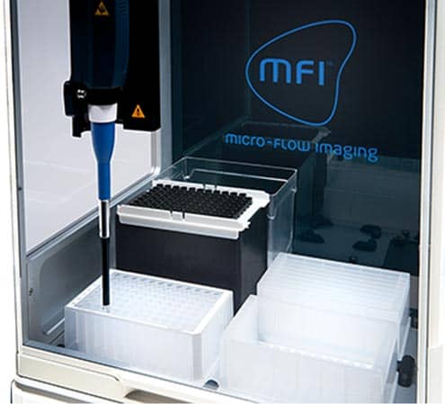
バイオ医薬品開発向けオートメーション: Bot1は、医薬品の製剤開発や製造プロセスを迅速かつ効率的に進めるために必要なスループットを提供します。1回の測定で、Bot1は最大90サンプルを手作業なしで処理できます。MFIとBot1は最大20センチポアズの粘度まで対応でき、最新のバイオ医薬品製剤が求める厳しい要件に対応します;製品を市場に投入するために必要な分析力を提供します。
解析を支援するソフトウェア
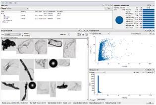
ユーザーを支援するソフトウェアソリューション: MFIソフトウェアにより、単一のサンプルを詳細に解析することも、複数サンプル全体を一度に評価することも可能です。MFIソフトウェアスイートは、使いやすいインターフェースで最大の分析力を発揮できるよう設計されたツール群を提供します。規制下での運用ですか?MFIソフトウェアは、21 CFR Part 11に準拠するために必要なすべての機能を備えています。
MFI 5000シリーズの参考文献
Micro-Flow Imaging(MFI)プラットフォームに関するすべての参考文献を見る
当社装置を使用した科学論文を集めた装置引用データベースで、関連論文をご覧ください。
Micro-Flow Imaging(MFI)をご利用のユーザーの声

「私たちはバイオシミラー企業として、自社製品が参照製剤と同等であるかを評価し、比較性評価や安定性試験にMFIを活用しています。さまざまな供給元からのサンプルを試験する必要がありますが、手動操作のみのシステムではスループットが不足するため、Bot1を備えた自動化MFIを採用しました。」
Micro-Flow Imaging+Bot1が支えるYuhwa Lee氏のバイオシミラー
Yuhwa Lee, Research Engineer, Samsung Bioepis

「MFIが提供する画像を用いることで、より詳細な解析を行い、このデータを他の手法と組み合わせて溶液中の粒子を迅速に識別・分類できます。これにより、クライアントは自社製品をより深く理解し、患者に最高品質の製品を提供していることを確認できます。」
Amber Fradkin氏、Micro-Flow Imaging(MFI)で製品品質の向上を実現
Amber Fradkin, Ph.D., Associate Director, Particle Characterization Core Facility, KBI Biopharma

「MFI技術により、治療用分子の安定性を高める条件をより深く理解できるようになりました。その結果、患者には世界水準の医薬品のみを届けることが可能になります。」
Stephanie Davies氏、MFIで治療用分子の製剤開発中の安定性プロファイルを完全把握
Stephanie Davies, Ph.D., Formulation Sciences, MedImmune
MFI 5000シリーズの詳細情報

MFIの仕組み
Micro-Flow Imagingは、デジタル顕微鏡による直接イメージングと、マイクロフルイディクスによる精密な流体制御を組み合わせた技術です。これにより、どのようなメリットが得られるのでしょうか? 85%のサンプリング効率を実現する高解像度画像により、試料中のすべてのサブビジブル粒子を対象に、形状情報を含めたより正確な粒子数測定およびサイズ評価が可能になります。さらに、タンパク質凝集体から気泡に至るまで、考えられるあらゆる粒子タイプを正確に識別できる、信頼性の高いデータを取得します。
サンプルがフローセルのセンサーゾーンを通過すると、画像が取得されます。取得されたすべての画像の粒子は個別に解析され、粒子数、サイズ、透過性、形態(形状)に関するデータベースが作成されます。さらに、画像はリアルタイムで表示されるため、その場で視覚的に確認できます。複数のサンプルの解析結果を同時に表示することも可能で、安定性や比較性のモニタリングも容易です。
| 項目 | MFI 5100 | MFI 5200 |
|---|---|---|
| 測定サイズ範囲 | 2~300 µm | 1~70 µm |
| サンプル解析率 | 85%以上(全サイズ範囲) | 85%以上(全サイズ範囲) |
| 焦点深度 (DOF) | 400 µm | 100 µm |
| フローセルの深さ | 400 µm (DOF一致) | 100 µm (DOF一致) |
| 解析流量 | 200 µL/分 | 150 µL/分 |
| 最大測定濃度 (@2.5 µm) | 175,000 粒子/mL | 900,000 粒子/mL |
| 自動化 | ハイスループットBot1オートサンプラー (オプション) | |
| サンプル導入方法 | ピペットチップ: 1 mL手動挿入、1 mLオートサンプラーシリンジバレル 2 mL、10 mL、20 mL手動挿入 BD Hypak™ Syringe導入アダプター: 1 mL、2 mL | |
| フローセルコーティング | 無コーティング、疎水性、カスタム | |
| 精密撹拌 | 可変速度制御: 200-2,000 RPM シリンジバレル対応 | |
| データ出力 | チャート形式: ヒストグラム、散布図、トレンドチャート 画像形式: 非圧縮TIFF(解析用)、圧縮JPG(保存用) | |
| 粒子計数パラメータ | 粒子数、濃度、質量、体積 | |
| 形態学パラメータ | サイズ(ECD)、最大フェレット径、アスペクト比、円形度、面積、周長、強度 | |
| カスタム解析フィルター | 2つ以上の画像・計数パラメータを組み合わせ可能、最大511種類のカスタム解析フィルター | |
| MFI View System Software (MVSS) 詳細は製品パンフレットをご覧ください | 手法ベースの解析プロトコル 時間分解解析とデータ圧縮による効率的な保存 21 CFR Part 11準拠機能 監査・ログ管理で安全にアクセス可能 MFI View Analysis Suite(MVAS)に完全対応 | |
| MFI View Analysis Suite (MVAS) 詳細は製品パンフレットをご覧ください | 画像、パラメータデータ、トレンドチャートの閲覧・解析 類似検索(Find-Similar)でサブグループを特定 マルチノードフィルターをリアルタイムで作成 データセットの重ね合わせと比較 複数カテゴリを含む詳細レポートの作成 繰り返し利用できる解析テンプレートの作成 | |
| 寸法 | MFI 5000単体: 幅 23.97 in × 奥行 17.01 in × 高さ 12.00 in Bot1オートサンプラー装着時: 幅 23.97 in × 奥行 17.01 in × 高さ 26.20 in | |
MFI 5000シリーズ製品資料
Bio-Techne.comでの日本語ページをご覧ください