
Human Pax2 Antibody
R&D Systems, part of Bio-Techne | Catalog # AF3364

Conjugate
Catalog #
Key Product Details
Species Reactivity
Validated:
Human
Cited:
Human, Mouse, Rat, Transgenic Mouse
Applications
Validated:
Immunohistochemistry, Western Blot
Cited:
Immunohistochemistry, Immunohistochemistry-Frozen, Western Blot, Flow Cytometry, Immunocytochemistry
Label
Unconjugated
Antibody Source
Polyclonal Goat IgG
Product Specifications
Immunogen
E. coli-derived recombinant human Pax2
Asp229-Pro363
Accession # Q02962
Asp229-Pro363
Accession # Q02962
Specificity
Detects human Pax2 in direct ELISAs and Western blots.
Clonality
Polyclonal
Host
Goat
Isotype
IgG
Scientific Data Images for Human Pax2 Antibody
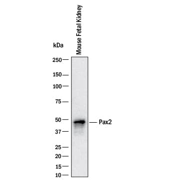
Detection of Mouse Pax2 by Western Blot.
Western blot shows lysates of mouse fetal kidney. PVDF Membrane was probed with 1 µg/mL of Goat Anti-Human Pax2 Antigen Affinity-purified Polyclonal Antibody (Catalog # AF3364) followed by HRP-conjugated Anti-Goat IgG Secondary Antibody (HAF017). A specific band was detected for Pax2 at approximately 47 kDa (as indicated). This experiment was conducted under reducing conditions and using Western Blot Buffer Group 1.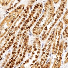
Pax2 in Human Kidney.
Pax2 was detected in immersion fixed paraffin-embedded sections of human kidney using Goat Anti-Human Pax2 Antigen Affinity-purified Polyclonal Antibody (Catalog # AF3364) at 1 µg/mL for 1 hour at room temperature followed by incubation with the Anti-Goat IgG VisUCyte™ HRP Polymer Antibody (VC004). Before incubation with the primary antibody, tissue was subjected to heat-induced epitope retrieval using Antigen Retrieval Reagent-Basic (CTS013). Tissue was stained using DAB (brown) and counterstained with hematoxylin (blue). Specific staining was localized to cell nuclei in convoluted tubules. Staining was performed using our protocol for IHC Staining with VisUCyte HRP Polymer Detection Reagents.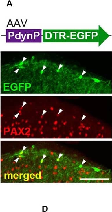
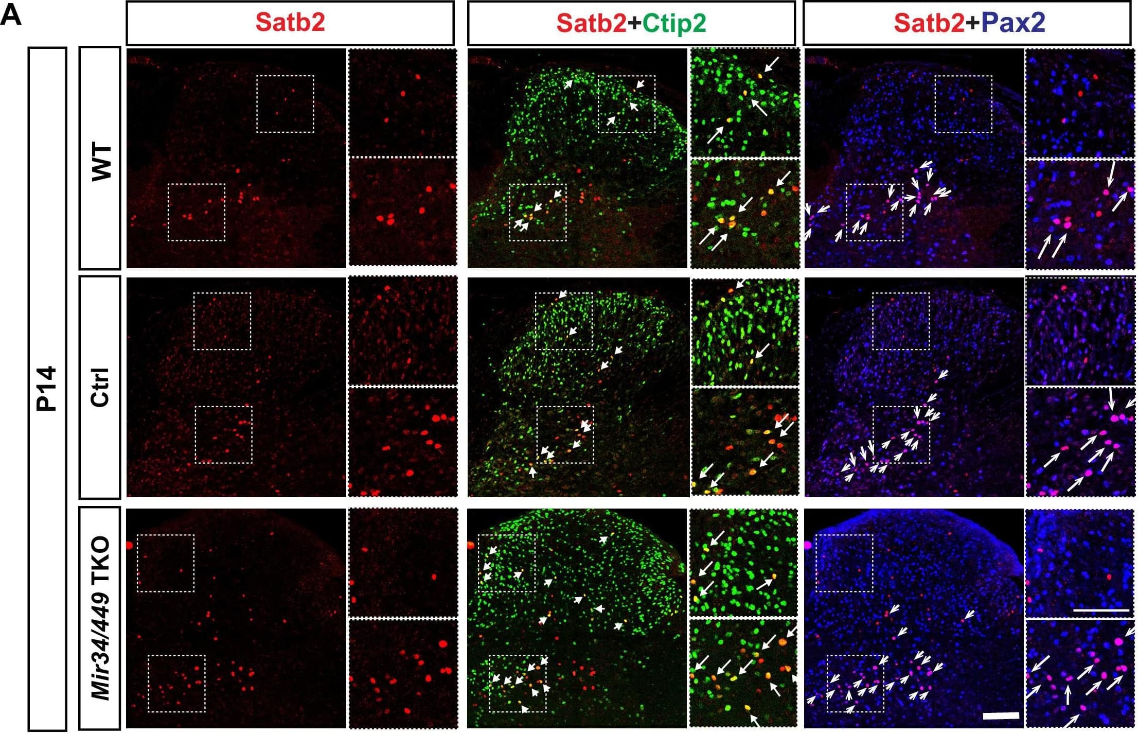
Detection of Rat Human Pax2 Antibody by Immunohistochemistry
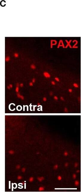
Ablation of AAV-PdynP+ spinal dorsal horn (SDH) neurons does not affect behavioral responses evoked by the optical stimulation of A beta fibers, nociceptive mechanical force, and heat in rats. (A) Enhanced green fluorescent protein (EGFP) expression (green) in the L4 SDH at 4 weeks after intra-SDH microinjection of AAV-PdynP-DTR-EGFP (indicated by an upper schematic illustration). Colocalization of EGFP and PAX2 (red) in the SDH (indicated by arrowheads). Scale bar, 100 μm. The percentage of co-expressing neurons was quantified (n = 3 rats tested, 94 total EGFP+ cells quantified, and 339 total PAX2+ cells quantified). (B) Diphtheria toxin receptors (DTR) immunofluorescence in the SDH after administration of diphtheria toxin (DTX: 50 μg/kg, i.p., two injection 72 h apart) or PBS. Scale bar, 200 μm. (C) Immunohistochemical analysis and quantification of PAX2+ neurons in the superficial laminae of the ipsilateral and contralateral SDH at 4 weeks after DTX injection (n = 6 rats tested, PAX2+ neurons: 992 [ipsi] and 1574 [contra] cells quantified). Scale bar, 50 μm. ****P < 0.0001, Unpaired t-test. (D) Paw withdrawal behaviors by light (score), von Frey filaments (threshold), and heat (latency) at 4 weeks after DTX or PBS injection (n = 7–9 rats). Data shown as mean ± SEM. Image collected and cropped by CiteAb from the following publication (https://pubmed.ncbi.nlm.nih.gov/35813063), licensed under a CC-BY license. Not internally tested by R&D Systems.
Applications for Human Pax2 Antibody
Application
Recommended Usage
Immunohistochemistry
1-15 µg/mL
Sample: Immersion fixed paraffin-embedded sections of human kidney
Sample: Immersion fixed paraffin-embedded sections of human kidney
Western Blot
1 µg/mL
Sample: Mouse fetal kidney
Sample: Mouse fetal kidney
Reviewed Applications
Read 2 reviews rated 4 using AF3364 in the following applications:
Formulation, Preparation, and Storage
Purification
Antigen Affinity-purified
Reconstitution
Reconstitute at 0.2 mg/mL in sterile PBS. For liquid material, refer to CoA for concentration.
Formulation
Lyophilized from a 0.2 μm filtered solution in PBS with Trehalose. See Certificate of Analysis for details.
*Small pack size (-SP) is supplied either lyophilized or as a 0.2 µm filtered solution in PBS.
*Small pack size (-SP) is supplied either lyophilized or as a 0.2 µm filtered solution in PBS.
Shipping
Lyophilized product is shipped at ambient temperature. Liquid small pack size (-SP) is shipped with polar packs. Upon receipt, store immediately at the temperature recommended below.
Stability & Storage
Use a manual defrost freezer and avoid repeated freeze-thaw cycles.
- 12 months from date of receipt, -20 to -70 °C as supplied.
- 1 month, 2 to 8 °C under sterile conditions after reconstitution.
- 6 months, -20 to -70 °C under sterile conditions after reconstitution.
Background: Pax2
Pax2 is a 40-45 kDa protein belonging to the small but developmentally important family of transcription regulators. Human Pax2 is a 416 amino acid (aa) residue protein with an N-terminal 128 aa DNA-binding paired box domain, a centrally-located octapeptide motif and a C-terminal truncated homeodomain. Based on the presence of the structural domains, Pax2 belongs to subgroup 2 in the Pax family. Pax2 is important for stem cell survival and lineage commitment during development. Pax2 is also expressed in various carcinomas where it seems to mediate anti-apoptotic functions. At least five splice isoforms of human Pax2 have been described within the region used as immunogen (shared by all isoforms). Human and mouse Pax2 share 98% aa sequence identity.
Long Name
Paired Box Gene 2
Alternate Names
paired box 2, paired box gene 2, paired box homeotic gene 2, paired box protein Pax-2
Gene Symbol
PAX2
UniProt
Additional Pax2 Products
Product Documents for Human Pax2 Antibody
Product Specific Notices for Human Pax2 Antibody
For research use only
Loading...
Loading...
Loading...
Loading...
Loading...