
Mouse F4/80 Antibody
R&D Systems, part of Bio-Techne | Catalog # MAB11811

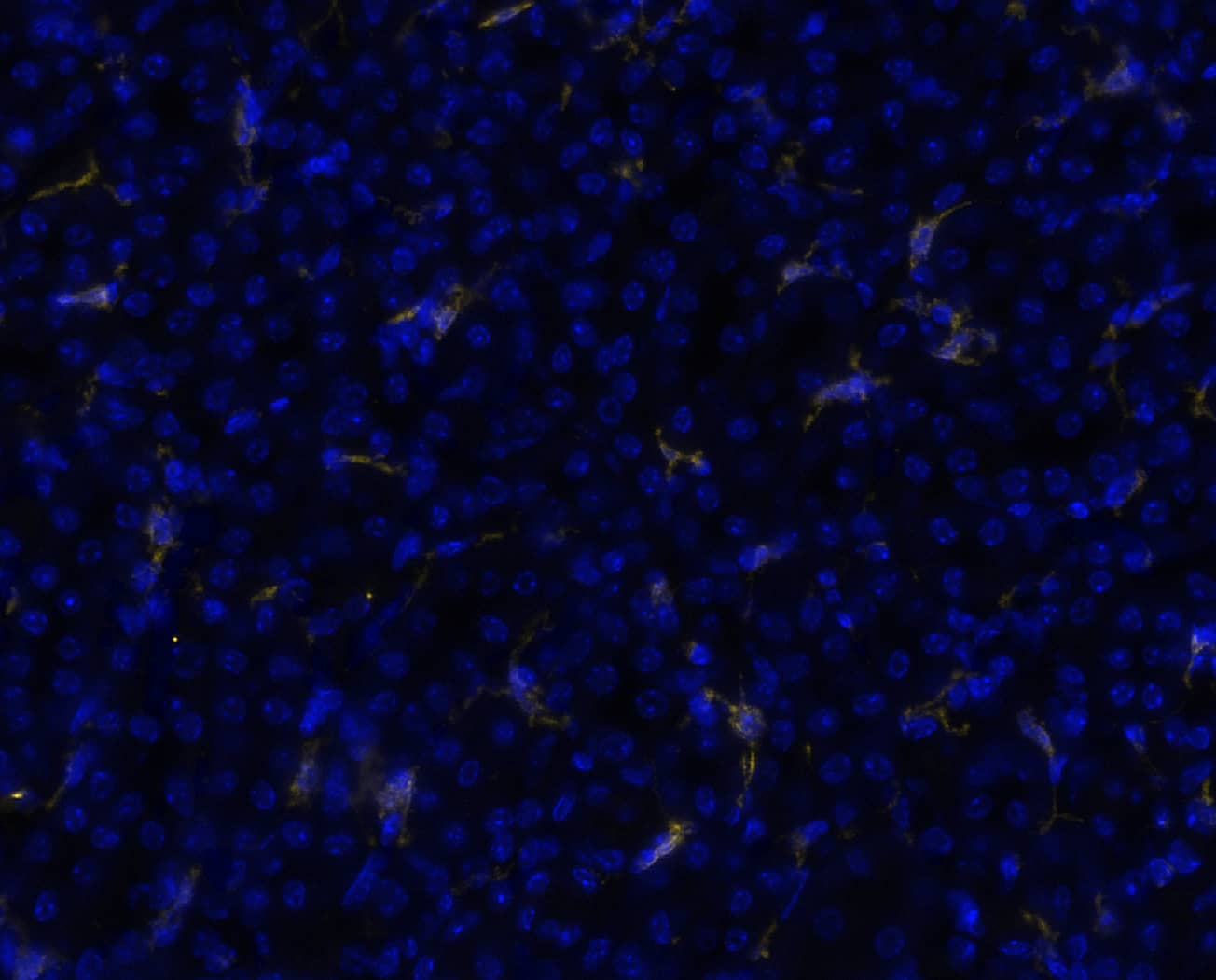
Key Product Details
Species Reactivity
Mouse
Applications
Immunohistochemistry, Western Blot, COMET
Label
Unconjugated
Antibody Source
Monoclonal Rat IgG2A Clone # 1123826
Product Specifications
Immunogen
Chinese Hamster Ovary cell line, CHO-derived mouse EMR-1
Met1-Ser590
Accession # Q61549
Met1-Ser590
Accession # Q61549
Specificity
Detects recombinant mouse F4/80 protein in Direct ELISA.
Clonality
Monoclonal
Host
Rat
Isotype
IgG2A
Scientific Data Images for Mouse F4/80 Antibody
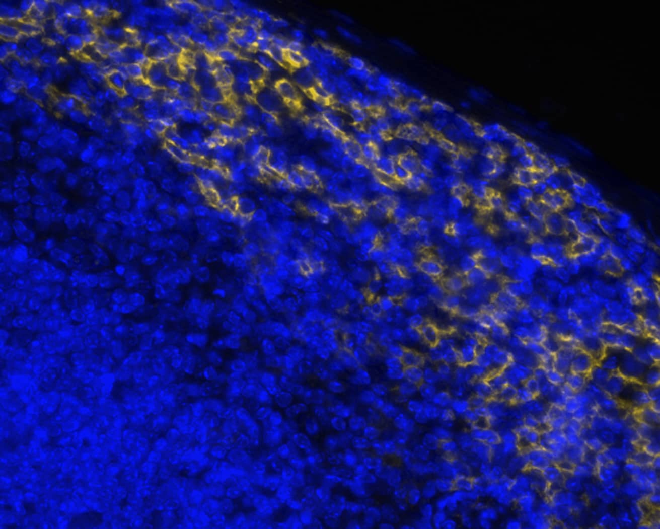
Detection of EMR1 in Mouse Spleen via seqIF™ staining on COMET™
EMR-1 was detected in immersion fixed paraffin-embedded sections of mouse spleen using Rat Anti-Mouse EMR-1 Monoclonal Antibody (Catalog # MAB11811) at 3ug/mL at 37 ° Celsius for 4 minutes. Before incubation with the primary antibody, tissue underwent an all-in-one dewaxing and antigen retrieval preprocessing using PreTreatment Module (PT Module) and Dewax and HIER Buffer H (pH 9; Epredia Catalog # TA-999-DHBH). Tissue was stained using the Alexa Fluor™ 647 Goat anti-Rat IgG Secondary Antibody at 1:200 at 37 ° Celsius for 2 minutes. (Yellow; Lunaphore Catalog # DR647RT) and counterstained with DAPI (blue; Lunaphore Catalog # DR100). Specific staining was localized to the membrane. Protocol available in COMET™ Panel Builder.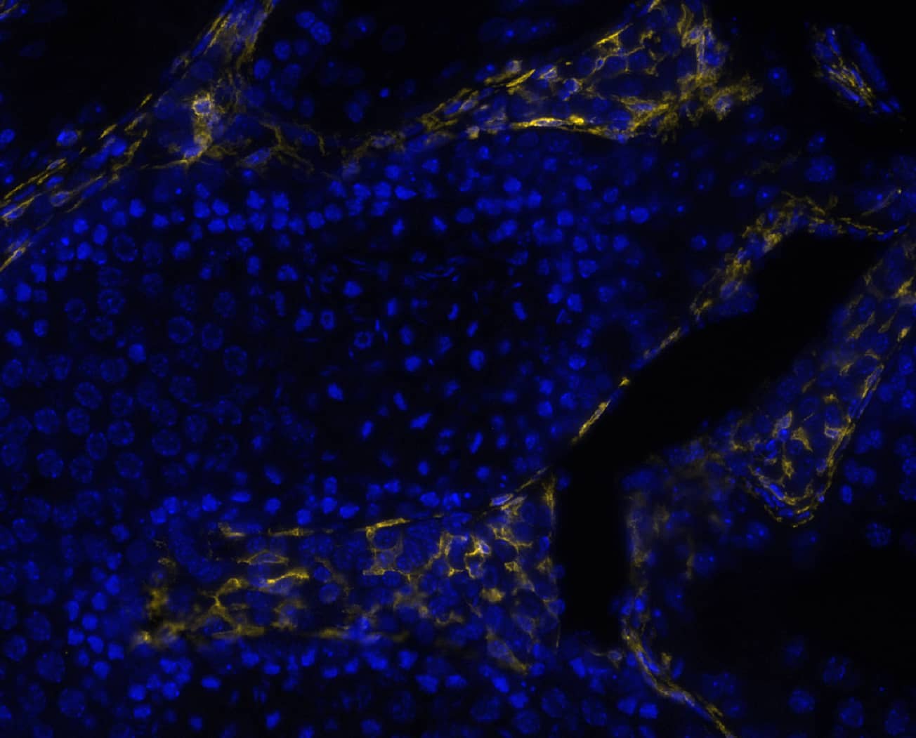
Detection of EMR1 in Mouse Testis via seqIF™staining on COMET™
EMR-1 was detected in immersion fixed paraffin-embedded sections of mouse testis using Rat Anti-Mouse EMR-1 Monoclonal Antibody (Catalog # MAB11811) at 3ug/mL at 37 ° Celsius for 4 minutes. Before incubation with the primary antibody, tissue underwent an all-in-one dewaxing and antigen retrieval preprocessing using PreTreatment Module (PT Module) and Dewax and HIER Buffer H (pH 9; Epredia Catalog # TA-999-DHBH). Tissue was stained using the Alexa Fluor™ 647 Goat anti-Rat IgG Secondary Antibody at 1:200 at 37 ° Celsius for 2 minutes. (Yellow; Lunaphore Catalog # DR647RT) and counterstained with DAPI (blue; Lunaphore Catalog # DR100). Specific staining was localized to the membrane. Protocol available in COMET™ Panel Builder.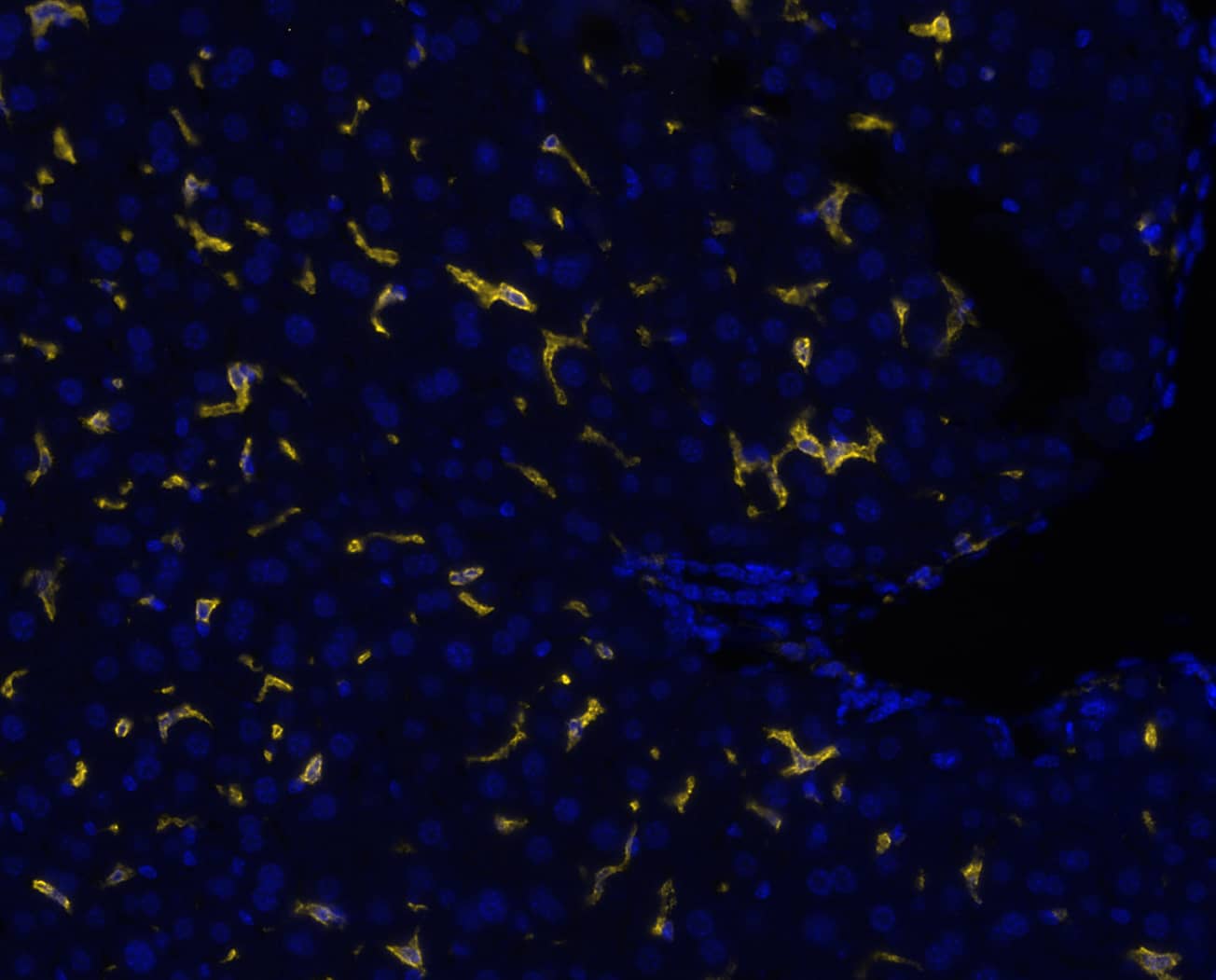
Detection of EMR1 in Mouse Liver via seqIF™staining on COMET™
EMR-1 was detected in immersion fixed paraffin-embedded sections of mouse liver using Rat Anti-Mouse EMR-1 Monoclonal Antibody (Catalog # MAB11811) at 3ug/mL at 37 ° Celsius for 4 minutes. Before incubation with the primary antibody, tissue underwent an all-in-one dewaxing and antigen retrieval preprocessing using PreTreatment Module (PT Module) and Dewax and HIER Buffer H (pH 9; Epredia Catalog # TA-999-DHBH). Tissue was stained using the Alexa Fluor™ 647 Goat anti-Rat IgG Secondary Antibody at 1:200 at 37 ° Celsius for 2 minutes. (Yellow; Lunaphore Catalog # DR647RT) and counterstained with DAPI (blue; Lunaphore Catalog # DR100). Specific staining was localized to the membrane. Protocol available in COMET™ Panel Builder.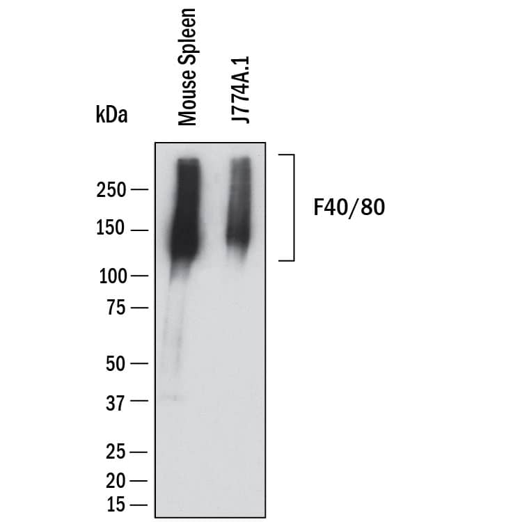
Applications for Mouse F4/80 Antibody
Application
Recommended Usage
COMET
3 µg/mL
Sample: Immersion fixed paraffin-embedded sections of mouse spleen, mouse testis, mouse liver and mouse kidney.
Sample: Immersion fixed paraffin-embedded sections of mouse spleen, mouse testis, mouse liver and mouse kidney.
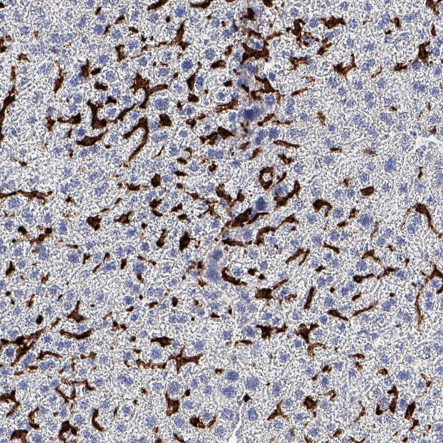
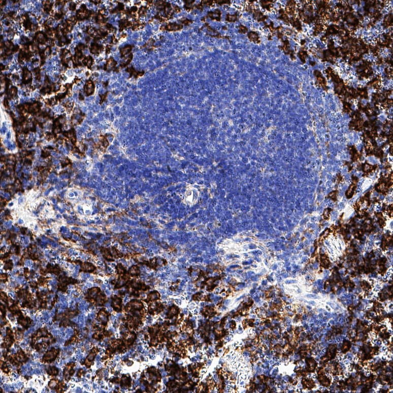
Immunohistochemistry
0.25-25 µg/mL
Sample: Perfusion fixed paraffin-embedded sections of mouse spleen and sections of mouse liver
Sample: Perfusion fixed paraffin-embedded sections of mouse spleen and sections of mouse liver
Western Blot
2 µg/mL
Sample: Mouse spleen and J774A.1 mouse reticulum cell sarcoma macrophage cell line
Sample: Mouse spleen and J774A.1 mouse reticulum cell sarcoma macrophage cell line
Formulation, Preparation, and Storage
Purification
Protein A or G purified from hybridoma culture supernatant
Reconstitution
Reconstitute lyophilized material at 0.2 mg/ml in sterile PBS. For liquid material, refer to CoA for concentration.
Formulation
Lyophilized from a 0.2 μm filtered solution in PBS with Trehalose.
Shipping
Lyophilized product is shipped at ambient temperature. Liquid small pack size (-SP) is shipped with polar packs. Upon receipt, store immediately at the temperature recommended below.
Stability & Storage
Use a manual defrost freezer and avoid repeated freeze-thaw cycles.
- 12 months from date of receipt, -20 to -70 °C as supplied.
- 1 month, 2 to 8 °C under sterile conditions after reconstitution.
- 6 months, -20 to -70 °C under sterile conditions after reconstitution.
Background: F4/80
Alternate Names
DD7A5-7, Gpf480, Ly71
Gene Symbol
ADGRE1
UniProt
Additional F4/80 Products
Product Documents for Mouse F4/80 Antibody
Product Specific Notices for Mouse F4/80 Antibody
For research use only
Loading...
Loading...
Loading...
Loading...
Loading...