Scientific Data Examples for Human HSPA8/HSC71 Antibody
Western Blot
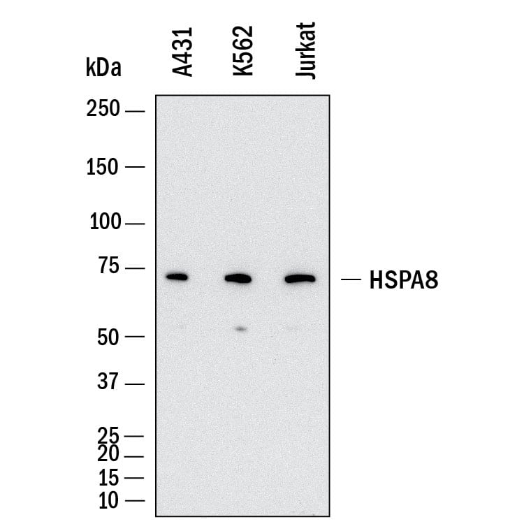
Detection of Human HSPA8/HSC71 by Western Blot.
Western Blot shows lysates of A431 human epithelial carcinoma cell line, K562 human chronic myelogenous leukemia cell line and Jurkat human acute T cell leukemia cell line. PVDF membrane was probed with 0.01 µg/ml of Rabbit Anti-Human HSPA8/HSC71 Monoclonal Antibody (Catalog # MAB11819) followed by HRP-conjugated Anti-Rabbit IgG Secondary Antibody (HAF008). A specific band was detected for HSPA8/HSC71 at approximately 70 kDa (as indicated). This experiment was conducted under reducing conditions and using Western Blot Buffer Group 1.
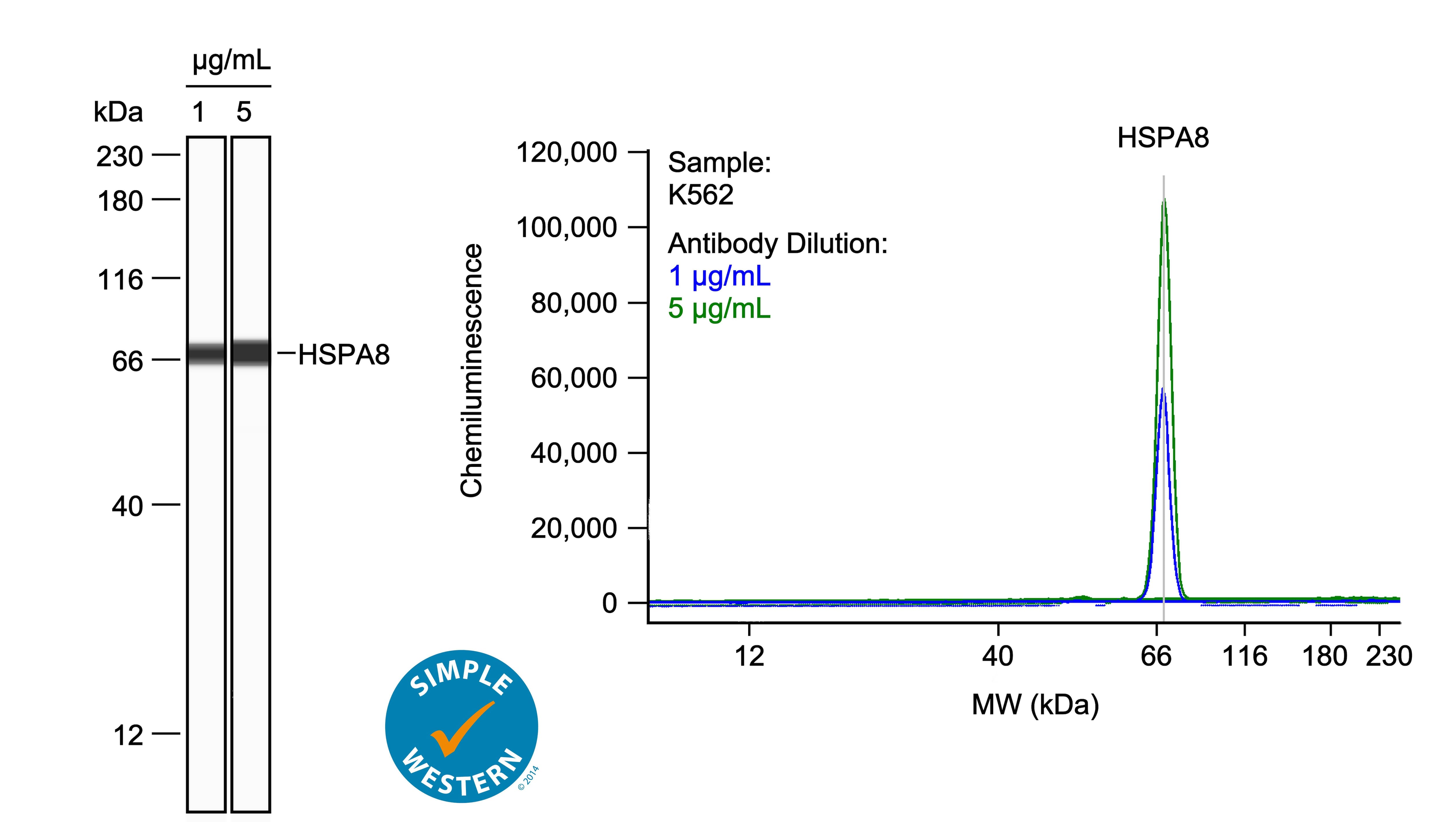
Simple Western
Detection of Human HSPA8/HSC71 by Simple WesternTM.Left: Simple Western lane view shows lysates of K562 human chronic myelogenous leukemia cell line, loaded at 0.1 mg/ml. A specific band was detected for HSPA8/HSC71 at approximately 70 kDa (as indicated) using both 1 µg/ml and 5 µg/ml of Rabbit Anti-Human HSPA8/HSC71 Monoclonal Antibody (Catalog # MAB11819) followed by HRP-conjugated Goat Anti-Rabbit Secondary Antibody (042-206). This experiment was conducted under reducing conditions and using the 12-230kDa separation system. Right: Simple Western electropherogram showing the same Rabbit Anti-Human HSPA8/HSC71 Monoclonal Antibody (Catalog # MAB11819) tested at 1 µg/ml (blue line) and 5 µg/ml (green line) in the K562 human chronic myelogenous leukemia cell line.
Immunohistochemistry
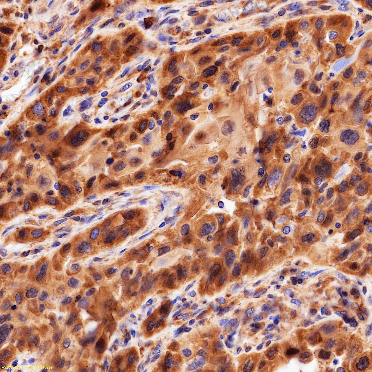
Immersion fixed paraffin-embedded sections of human lung
HSPA8/HSC71 was detected in immersion fixed paraffin-embedded sections of human lung using Rabbit Anti-Human HSPA8/HSC71 Monoclonal Antibody (Catalog # MAB11819) at 1 µg/ml overnight at 4 °C. Before incubation with the primary antibody, tissue was subjected to heat-induced epitope retrieval using VisUCyte Antigen Retrieval Reagent-Basic (VCTS021). Tissue was stained using the HRP-conjugated Anti-Rabbit IgG Secondary Antibody (HAF008) and counterstained with hematoxylin (blue). Specific staining was localized to the cytoplasm and nucleus. View our protocol for Chromogenic IHC Staining of Paraffin-embedded Tissue Sections.
Formulation, Preparation and Storage
Purification
Protein A or G purified from hybridoma culture supernatant
Reconstitution
Reconstitute lyophilized material at 0.2 mg/ml in sterile PBS. For liquid material, refer to CoA for concentration.
Formulation
Lyophilized from a 0.2 μm filtered solution in PBS with Trehalose.
Shipping
Lyophilized product is shipped at ambient temperature. Liquid small pack size (-SP) is shipped with polar packs. Upon receipt, store immediately at the temperature recommended below.
Stability & Storage
Use a manual defrost freezer and avoid repeated freeze-thaw cycles.
12 months from date of receipt, -20 to -70 °C as supplied.
1 month, 2 to 8 °C under sterile conditions after reconstitution.
6 months, -20 to -70 °C under sterile conditions after reconstitution.
Reconstitution Calculator
Background: HSPA8/HSC71
Heat shock cognate 71-kDa protein (HSPA8), also referred to as HSC70, is a member of the heat shock protein 70 (HSP70) family, with a molecular weight of approximately 71 kDa. HSPA8 is an ATP-dependent molecular chaperone ubiquitously expressed in eukaryotic cells and plays a critical role in protein homeostasis. It facilitates the folding of nascent polypeptides, the assembly and disassembly of protein complexes, and the translocation of proteins across membranes. Additionally, HSPA8 is involved in the targeting and degradation of misfolded or damaged proteins via the ubiquitin-proteasome pathway and lysosomal-associated degradation. Beyond its canonical chaperone functions, HSPA8 participates in cellular processes such as autophagy, stress response, and clathrin-mediated endocytosis. Dysregulation of HSPA8 expression or activity has been implicated in various pathological conditions, including neurodegenerative disorders such as Alzheimer's and Parkinson's diseases, where aberrant protein aggregation is a hallmark feature. The multifunctionality and essential cellular roles of HSPA8 underscore its utility as both a biomarker and a potential therapeutic target in a range of diseases.
Item
Value
References
Hartl FU, Hayer-Hartl M. Molecular chaperones in the cytosol: from nascent chain to folded protein. Science. 2002 Mar 8;295(5561):1852-8. doi: 10.1126/science.1068408. PMID: 11884745.
Pustovaya K, Venediktov A, Soldatov V, Kuzmin E, Pokidova K, Gartzeva V, Payushina O, Tsytsarev V, Meglinski I and Piavchenko G (2026) Recent insights into HSP70: proteostasis and beyond. Front. Mol. Biosci. 13:1791536. doi: 10.3389/fmolb.2026.1791536.
Long Name
Heat Shock 70 kDa Protein 8
Entrez Gene IDs
3312 (Human)
Alternate Names
HSC54, HSC70, HSC70heat shock 70kd protein 10, HSC71, HSP71, HSP73, HSP73constitutive heat shock protein 70, HSPA10, HSPA8, Heat shock 70 kDa protein 8, LAP1, LPS-associated protein 1, MGC131511, MGC29929, N-myristoyltransferase inhibitor protein 71, NIP71, heat shock 70kD protein 8, heat shock 70kDa protein 8, heat shock cognate 71 kDa protein, heat shock cognate protein 54, heat shock cognate protein, 71-kDa, lipopolysaccharide-associated protein 1