MauriceFlex: Flex Your Freedom

Meet MauriceFlex

Meet MauriceFlex, your fully integrated capillary electrophoresis platform that gives you:

  • Automated imaged icIEF and CE-SDS assays
  • Same-day charge variant fraction collection
  • Flexibility to analyze fractions with intact mass, reduced mass, peptide mapping, and others
  • 21CFR Part 11 compliance for your data

MauriceFlex combines protein charge isoform fractionation with imaged-capillary isoelectric focusing (icIEF) and CE-SDS analysis in one instrument, so that you can go beyond routine size and charge and conduct an in-depth analysis of your molecule at any stage without needing to rely on different instruments, methods, or expertise.

Charge isoform separation and peak mobilization data from Maurice Flex
A.icIEF profiles of charge isoforms. B. Mobilization of charge isoform peaks for fraction collection
Imaged cief protein fractionation on Maurice Flex - banner

Why Fractionate Proteins With MauriceFlex?

The potency, safety, and efficacy of your biotherapeutic can be impacted by certain charge variants present, therefore in-depth characterization is crucial. Now, instead of spending weeks or months on ion-exchange chromatography (IEX) based fractionation, detect charge variants of your protein, AND collect their fractions on the same instrument in the same day!  

MauriceFlex Gives You Freedom

  • Freedom to do more with your fractions – analyze with intact mass, subunit, peptide mapping, and more
  • Freedom from laborious IEX workflow – easily collect high-purity fractions without needing columns
  • Freedom from undesirable clones – implement fractionation in early discovery stages
  • Freedom to do more with your time - identify, separate, and fractionate charge isoforms in less than a day
  • Freedom to do more with your budget – get the maximum value out of a single instrument

When to Fractionate With MauriceFlex

  • Biopharma discovery/Cell culture – Without spending time and effort on IEX, get valuable information on PTMs responsible for the charge isoforms so you can select the right clone for your molecule
  • Analytical development – Conduct routine CE-SDS and icIEF assays, and collect icIEF fractions on the same instrument without needing to develop additional methods
  • Formulation – Identify various PTMs impacting the stability of your molecule in less than day

icIEF Fractionation Workflow With MauriceFlex

Maurice Flex Workflow - 6 steps for protein fractionation based on ic-ief

How icIEF Fractionation Works

  • An applied electric field creates a pH gradient across the capillary inside the MauriceFlex cartridge
  • Proteins with different isoelectric points or pI values migrate along the capillary until they reach pH values matching their pI, leading to focusing and charge separation
  • For fractionation, the catholyte end of the capillary is brought in contact with ammonium acetate
    The applied voltage drives the acetate ions into the capillary from the catholyte end and the proton from the anolyte end
  • The displacement of hydroxide ions by acetate ions gradually changes the pH gradient along the capillary, mobilizing the previously focused proteins to move along the new pH gradient towards the cathodic end 
  • Proteins elute out of the capillary one by one while leaving urea and other impurities behind, giving you high-purity fractions you need for further analysis
MauriceFlex Fractionation Workflow

Advantages of icIEF Fractionation with MauriceFlex

MauriceFlex Advantages
Perform Get Use In Analyze Further With
  • CE-SDS
  • icIEF
  • icIEF fraction collection
  • Same-day method development
  • >80% purity of fractions
  • Ability to pool fractions
  • Early discovery
  • Analytical Development
  • Formulation
  • Intact mass, reduced mass
  • Peptide mapping


MauriceFlex Instrument performs ic-ief based protein fractionation

Charge variant fraction collection with MauriceFlex renders significant advantages on time and capital when compared with IEX and other existing technologies. The key advantages are:

  • Same-day fraction collection
  • High purity fractions
  • Single fractionation offering enough material for intact MS analysis
  • Compatibility with urea and methylcellulose
  • Ability to pool fractions for peptide mapping
  • Same system offering CE-SDS and cIEF assays
  • Eliminating the need for bridging studies

Featured Resources

Press Release for protein fractionation on MauriceFlex Teaser Card

Press Release

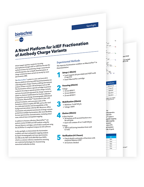
MauriceFlex™ icIEF Analysis Adds Fractionation, Enabling Mass Spec Peak Characterization

c-ief protein fractionation with MauriceFlex Poster Teaser Card

Scientific Poster

Novel icIEF Fractionation Coupled with Different MS Systems for Rapid Charge Variant Characterization of Therapeutic Antibodies

Which Maurice is right for you?


System

Maurice

Maurice C.

Maurice S.

Maurice Flex

icIEF Charge Application

Yes

Yes

No

Yes

CE-SDS Size Application

Yes

No

Yes

Yes
icIEF Fractionation No No No Yes

Absorbance Detection

Yes

Yes

Yes

Yes

Fluorescence Detection

Yes

Yes

No

Yes

Onboard Mixing for Sample Prep

Yes

Yes

No

No

Specifications

Description

cIEF

CE-SDS PLUS

Turbo CE-SDS

cIEF Fractionation

Minimum Sample Volume

50 µL

50 µL

100 µL

100 µL

Sample Delivery

Vacuum

Electrokinetic

Electrokinetic

Vacuum

Typical Separation Time

6-10 min (molecule-dependent)

Reduced IgG: 25 minutes; Non-reduced IgG: 35 minutes

Reduced IgG: 5.5 minutes; Non-reduced IgG: 8 minutes

40 – 50 min (molecule-dependent)
Detection Capability UV Absorbance at 280 nm

Fluorescence: Ex 280 nm, Em 320–450 nm
UV Absorbance at 220 nm UV Absorbance at 220 nm Fluorescence: Ex 280 nm, Em 320-450nm

Typical Voltage

Pre-focusing: 1500 V
Focusing: 3000 V

Separation: 5750 V

Separation: 4,200 V

Pre-focusing: 500 V and 1000 V
Focusing: 1500 V

Sample Injections per Cartridge

100 guaranteed, 200 maximum (max 25 batches)

100 guaranteed, 500 maximum (max 25 batches)

100 guaranteed (max 25 batches)

Maximum 15 injections

Maximum Sample Injections per Batch

100

48

96

1 (fractionation)
4 (cIEF)
 

pI/Size Range

2.85-10.45

10-270 kDa

10-270 kDa

3-10

pI/Sizing CV

1%

≤2%

<2%

1%

CV for Peaks >10% Composition

≤5% (Intra-batch), ≤6% (Inter-batch)

N/A

N/A

≤10% (Inter-batch)
Relative Migration Time CV N/A <1% for reduced IgG <5% N/A

pI/Sizing Resolution

0.05 pl units (for wide range 3-10 ampholyte)

≥ 1.5 for NGHC/HC IgG Standard)

≥ 1.0 for NGHC/HC IgG Standard)

N/A

Dynamic Range

2 logs

2 logs

2 logs

N/A

Linearity

>0.995

>0.995

>0.995

N/A

Sensitivity (LOD)

0.7 µg/mL (Native fluorescence)
3.0 µg/mL (Absorbance) (Values based on a monoclonal antibody)

0.3 µg/mL
(Value based on internal standard)

0.6 μg/mL (value based on internal standard)

N/A

Sample Tray Options

96-well plates or 48 vials

96-well plates or 48 vials

96-well plates or 48 vials

96-well plates only

Power

100 V-240 V (AC), 50/60 Hz, 500 W

100 V-240 V (AC), 50/60 Hz, 500 W

100 V-240 V (AC), 50/60 Hz, 500 W

100 V-240 V (AC), 50/60 Hz, 500 W
Voltage Range 0–6,500 V 0–6,500 V 0–6,500 V 0–6,500 V

Temperature Control Range

4-25 °C

4-25 °C

4-25 °C

10-25 °C

Dimensions

44cm H x 42cm W x 61cm D

44cm H x 42cm W x 61cm D

44cm H x 42cm W x 61cm D

44cm H x 42cm W x 61cm D

Weight

46 kg (100 lb)

46 kg (100 lb)

46 kg (100 lb)

46 kg (100 lb)