
Human IL-17E/IL-25 Antibody
R&D Systems, part of Bio-Techne | Catalog # MAB1258


Scientific images may be resized, cropped, or adjusted for brightness or contrast for presentation purposes. These updates are limited to presentation and do not affect the underlying scientific interpretation of the data. Where available, additional source data may be provided upon request.
Conjugate
Catalog #
Key Product Details
Species Reactivity
Validated:
Human
Cited:
Human, Mouse
Applications
Validated:
Western Blot, ELISA, Intracellular Staining by Flow Cytometry, CyTOF-ready
Cited:
Immunohistochemistry, Immunohistochemistry-Paraffin, Western Blot, Flow Cytometry, Immunocytochemistry
Label
Unconjugated
Antibody Source
Monoclonal Mouse IgG1 Clone # 182203
Product Specifications
Immunogen
E. coli-derived recombinant human IL-17E
Tyr33-Gly177
Accession # Q9H293
Tyr33-Gly177
Accession # Q9H293
Specificity
Detects human IL-17E in direct ELISAs and Western blots. Shows 100% cross-reactivity with recombinant mouse IL‑17E and no cross-reactivity with recombinant human (rh) IL-17, rhIL-17B, rhIL-17C, or rhIL-17F.
Clonality
Monoclonal
Host
Mouse
Isotype
IgG1
Scientific Data Images for Human IL-17E/IL-25 Antibody
Detection of IL-17E/IL-25 in PC-3 Human Cell Line by Flow Cytometry.
PC-3 human prostate cancer cell line was stained with Human IL-17E/IL-25 Monoclonal Antibody (Catalog # MAB1258, filled histogram) or isotype control antibody (Catalog # MAB002, open histogram), followed by Allophycocyanin-conjugated Anti-Mouse IgG F(ab')2Secondary Antibody (Catalog # F0101B). To facilitate intracellular staining, cells were fixed with paraformaldehyde and permeabilized with saponin.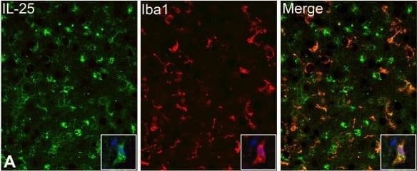
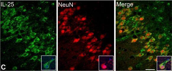
Detection of Human IL-17E/IL-25 by Immunocytochemistry/Immunofluorescence
IL-25 IR in neurons, astrocytes and microglia. IFC reveals IL-25 IR (in green) co-localizing with three CNS cell type markers (red), (A) Iba1, a microglial marker, (B) GFAP, an astrocytic marker and (C) NeuN, a neuronal marker. Panel A,B: CA4; Panel C: dentate gyrus. Note that IL-25 IR is also present in the astrocytic endfeet surrounding the bloodvessels (B). The insets are higher power magnifications taken from a representative area, with the added nuclear marker DAPI (blue). Scale bar = 40 μm Image collected and cropped by CiteAb from the following publication (https://pubmed.ncbi.nlm.nih.gov/22935090), licensed under a CC-BY license. Not internally tested by R&D Systems.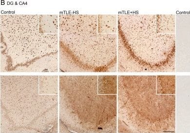
Detection of Human IL-17E/IL-25 by Immunocytochemistry/Immunofluorescence
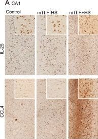
Hippocampal expression patterns of IL-25 and CCL4 in Control, mTLE-HS and mTLE + HS patients. Photomicrographs showing typical examples of Il-25 and CCL4 staining in the hippocampal CA1 region (A) and the DG/CA4 region (B). IL-25 immunoreactivity (IR) is evident in cells with neuronal morphology in all three patient groups (A, B); note the increased IL-25 staining in the mTLE + HS hippocampus in small cells. CCL4 IR is low in controls (A, B). Increased CCL4 IR is detected in both mTLE patient groups in the DG-CA4 area (B) and in the CA1 area of mTLE + HS patients (A). Scale bar = 200 μm.The insets are higher power magnifications taken from the same anatomical area Image collected and cropped by CiteAb from the following publication (https://pubmed.ncbi.nlm.nih.gov/22935090), licensed under a CC-BY license. Not internally tested by R&D Systems.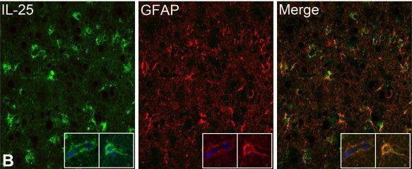
Applications for Human IL-17E/IL-25 Antibody
Application
Recommended Usage
CyTOF-ready
Ready to be labeled using established conjugation methods. No BSA or other carrier proteins that could interfere with conjugation.
ELISA
This antibody functions as an ELISA capture antibody when paired with Goat Anti-Human IL‑17E/IL‑25 Antigen Affinity-purified Polyclonal Antibody (Catalog # AF1258).
This product is intended for assay development on various assay platforms requiring antibody pairs.
Intracellular Staining by Flow Cytometry
2.5 µg/106 cells
Sample: PC-3 human prostate cancer cell line fixed with paraformaldehyde and permeabilized with saponin
Sample: PC-3 human prostate cancer cell line fixed with paraformaldehyde and permeabilized with saponin
Western Blot
1 µg/mL
Sample: Recombinant Human IL-17E/IL-25 (Catalog # 1258-IL)
Sample: Recombinant Human IL-17E/IL-25 (Catalog # 1258-IL)
Reviewed Applications
Read 1 review rated 5 using MAB1258 in the following applications:
Formulation, Preparation, and Storage
Purification
Protein A or G purified from hybridoma culture supernatant
Reconstitution
Reconstitute at 0.5 mg/mL in sterile PBS. For liquid material, refer to CoA for concentration.
Formulation
Lyophilized from a 0.2 μm filtered solution in PBS with Trehalose. *Small pack size (SP) is supplied either lyophilized or as a 0.2 µm filtered solution in PBS.
Shipping
Lyophilized product is shipped at ambient temperature. Liquid small pack size (-SP) is shipped with polar packs. Upon receipt, store immediately at the temperature recommended below.
Stability & Storage
Use a manual defrost freezer and avoid repeated freeze-thaw cycles.
- 12 months from date of receipt, -20 to -70 °C as supplied.
- 1 month, 2 to 8 °C under sterile conditions after reconstitution.
- 6 months, -20 to -70 °C under sterile conditions after reconstitution.
Background: IL-17E/IL-25
The Interleukin 17 (IL-17) family proteins, comprising six members (IL-17, and IL-17B through IL-17F), are secreted, structurally related proteins that share a conserved cysteine-knot fold near the C-terminus, but have considerable sequence divergence at the N-terminus. With the exception of IL-17B, which exists as a non-covalently linked dimer, all IL-17 family members are disulfide-linked dimers. IL-17 family proteins are pro-inflammatory cytokines that induce local cytokine production and are involved in the regulation of immune functions (1, 2).
Human IL-17E cDNA encodes a 177 amino acid (aa) residues precursor protein with a putative 32 aa signal peptide (3). A second isoform of human IL-17E encoding a 161 aa precursor protein also exists (4). The two isoforms differ in their signal peptide sequences. Mature human IL-17E shares 76% aa sequence identity with mature mouse IL-17E. Human IL-17E also shares from 25-36% aa sequence identity with the other human IL-17 family members. IL-17E expression was detected at very low levels by PCR in various peripheral tissues including brain, kidney, lung, prostate, testis, adrenal gland, spinal cord, and trachea (3). IL-17E binds and activates IL-17 B Receptor (IL-17B R) (alternatively known as IL-17 Rh1, IL-17E R, and EVI27) (3), which is expressed in kidney and liver, and at lower levels in brain, testis, and other endocrine tissues. The expression of IL-17B R is up regulated under inflammatory conditions. Ligation of IL-17E to IL-17 RB induces activation of nuclear factor kappa-B and stimulates the production of the pro-inflamatory cytokine IL-8 (3). IL-17 has also been found to promote the expression of the prototypical Th2 genes (4, 5).
References
- Aggarwal, S. and A.L. Gurney (2002) J. Leukoc. Biol. 71:1.
- Moseley, T.A. et al. (2003) Cytokine & Growth Factor Rev. 14:155.
- Lee, J. et al. (2001) J. Biol. Chem. 276:1660.
- Hurst, S.D. et al. (2002) J. Immunol. 169:443.
- Pan, G. et al. (2001) J. Immunol. 167:6569.
Long Name
Interleukin 17E
Alternate Names
IL-25, IL17E, IL25
Gene Symbol
IL25
UniProt
Additional IL-17E/IL-25 Products
Product Documents for Human IL-17E/IL-25 Antibody
Product Specific Notices for Human IL-17E/IL-25 Antibody
For research use only
Loading...
Loading...
Loading...
Loading...
Loading...
Loading...