
Human TIM-4 Antibody
R&D Systems, part of Bio-Techne | Catalog # AF2929



Scientific images may be resized, cropped, or adjusted for brightness or contrast for presentation purposes. These updates are limited to presentation and do not affect the underlying scientific interpretation of the data. Where available, additional source data may be provided upon request.
Conjugate
Catalog #
Key Product Details
Species Reactivity
Validated:
Human
Cited:
Human
Applications
Validated:
Immunohistochemistry, Western Blot
Cited:
Immunohistochemistry-Paraffin, Neutralization, Flow Cytometry, Immunocytochemistry
Label
Unconjugated
Antibody Source
Polyclonal Goat IgG
Product Specifications
Immunogen
Mouse myeloma cell line NS0-derived recombinant human TIM-4 (R&D Systems, Catalog # 2929-TM)
Glu25-Leu315
Accession # Q96H15
Glu25-Leu315
Accession # Q96H15
Specificity
Detects human TIM-4 in direct ELISAs and Western blots. In direct ELISAs, less than 1% cross‑reactivity with recombinant human (rh) TIM-1 and recombinant mouse (rm) TIM-4 is observed.
Clonality
Polyclonal
Host
Goat
Isotype
IgG
Scientific Data Images for Human TIM-4 Antibody
Detection of Recombinant Human TIM‑4 by Western Blot.
Western blot shows 25 ng of Recombinant Human TIM-4 His-tag (Catalog # 2929-TM), Recombinant Human TIM-1/KIM-1/HAVCR (Catalog # 1750-TM), Recombinant Human TIM-3 Fc Chimera (Catalog # 2365-TM), Recombinant Mouse TIM-1/KIM-1/HAVCR (Catalog # 1817-TM), Recombinant Mouse TIM-2 (Catalog # 1885-TM), Recombinant Mouse TIM-3 Fc Chimera (Catalog # 1529-TM), Recombinant Mouse TIM-4 (Catalog # 2826-TI), Recombinant Mouse TIM-5, and Recombinant Mouse TIM-6. PVDF Membrane was probed with 0.1 µg/mL of Goat Anti-Human TIM-4 Antigen Affinity-purified Polyclonal Antibody (Catalog # AF2929) followed by HRP-conjugated Anti-Goat IgG Secondary Antibody (Catalog # HAF109). A specific band was detected for TIM-4 at approximately 75 kDa (as indicated). This experiment was conducted under reducing conditions and using Immunoblot Buffer Group 3.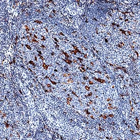
TIM‑4 in Human Tonsil.
TIM-4 was detected in immersion fixed paraffin-embedded sections of human tonsil using Goat Anti-Human TIM-4 Antigen Affinity-purified Polyclonal Antibody (Catalog # AF2929) at 1 µg/mL for 1 hour at room temperature followed by incubation with the Anti-Goat IgG VisUCyte™ HRP Polymer Antibody (Catalog # VC004). Before incubation with the primary antibody, tissue was subjected to heat-induced epitope retrieval using Antigen Retrieval Reagent-Basic (Catalog # CTS013). Tissue was stained using DAB (brown) and counterstained with hematoxylin (blue). Specific staining was localized to cell membranes of macrophages. View our protocol for IHC Staining with VisUCyte HRP Polymer Detection Reagents.
Detection of TIM-4 by Immunohistochemistry
Double F-IHC for TIM4 and surface markers of immune cells in breast carcinoma tissues. Double F-IHC staining was performed for TIM4 (green) and CD68 (a, red), CD11c (b, red), and CD20 (c, red), with DAPI (blue) used for nuclear counterstaining, in human breast carcinoma tissues. Human lymph node tissue was used as a positive control for TIM4 (d), CD68 (e), CD11c (f), and CD20 (g) staining. All images were acquired at 400 × magnification Image collected and cropped by CiteAb from the following open publication (https://pubmed.ncbi.nlm.nih.gov/41563642), licensed under a CC-BY license. Not internally tested by R&D Systems.


Applications for Human TIM-4 Antibody
Application
Recommended Usage
Immunohistochemistry
1-15 µg/mL
Sample: Immersion fixed paraffin-embedded sections of human tonsil
Sample: Immersion fixed paraffin-embedded sections of human tonsil
Western Blot
0.1 µg/mL
Sample: Recombinant Human TIM-4 (Catalog # 2929-TM)
Sample: Recombinant Human TIM-4 (Catalog # 2929-TM)
Reviewed Applications
Read 1 review rated 4 using AF2929 in the following applications:
Formulation, Preparation, and Storage
Purification
Antigen Affinity-purified
Reconstitution
Reconstitute at 0.2 mg/mL in sterile PBS. For liquid material, refer to CoA for concentration.
Formulation
Lyophilized from a 0.2 μm filtered solution in PBS with Trehalose. *Small pack size (SP) is supplied either lyophilized or as a 0.2 µm filtered solution in PBS.
Shipping
Lyophilized product is shipped at ambient temperature. Liquid small pack size (-SP) is shipped with polar packs. Upon receipt, store immediately at the temperature recommended below.
Stability & Storage
Use a manual defrost freezer and avoid repeated freeze-thaw cycles.
- 12 months from date of receipt, -20 to -70 °C as supplied.
- 1 month, 2 to 8 °C under sterile conditions after reconstitution.
- 6 months, -20 to -70 °C under sterile conditions after reconstitution.
Background: TIM-4
TIM-4 (T cell; immunoglobulin; mucin-4), also known as SMUCKLER, is a 60 kDa member of the TIM family of immune regulating proteins. TIMs are type I transmembrane proteins with one Ig-like V domain and one Ser/Thr-rich mucin domain (1 - 3). The human TIM-4 cDNA encodes a 378 amino acid (aa) precursor that includes a 24 aa signal sequence, a 290 aa extracellular domain (ECD), a 21 aa transmembrane segment, and a 43 aa cytoplasmic tail (4). Structurally, TIM-4 is distinguished from other TIMs by the presence of an RGD motif in its Ig domain and the lack of a site for tyrosine phosphorylation in its cytoplasmic tail. The mucin domain in TIM-4 is larger than in TIM-1 or TIM-3. Within the ECD, human TIM-4 shares 35% and 23% aa sequence identity with TIM-1 and TIM-3, respectively. A TIM-2 ortholog has not been identified in human. The ECD of human TIM-4 shares 45% aa sequence identity with that of mouse and rat TIM-4. TIM-4 is expressed by macrophages and mature dendritic cells but not by lymphocytes (4, 5). TIM-4 binds specifically to TIM-1 which is also the cellular receptor for the hepatitis A virus, and has been implicated in the development of asthma (5 - 7). Among hematopoietic cells, TIM-1 is expressed on activated B and T cells, preferentially in the Th2 subset of CD4+ T cells (5, 8). The interaction of TIM-4 with TIM-1 induces costimulatory and hyperproliferative signals in T cells (5).
References
- Kuchroo, V.K. et al. (2003) Nat. Rev. Immunol. 3:454.
- Mariat, C. et al. (2005) Phil. Trans. R. Soc. B 360:1681.
- Meyers, J.H. et al. (2005) Trends Mol. Med. 11:362.
- Shakhov, A.N. et al. (2004) Eur. J. Immunol. 34:494.
- Meyers, J.H. et al. (2005) Nat. Immunol. 6:455.
- Feigelstock, D. et al. (1998) J. Virol. 72:6621.
- McIntire, J.J. et al. (2001) Nat. Immunol. 2:1109.
- Khademi, M. et al. (2004) J. Immunol. 172:7169.
Long Name
T Cell Immunoglobulin Mucin-4
Alternate Names
Smuckler, TIM4, TIMD4
Gene Symbol
TIMD4
UniProt
Additional TIM-4 Products
Product Documents for Human TIM-4 Antibody
Product Specific Notices for Human TIM-4 Antibody
For research use only
Loading...
Loading...
Loading...
Loading...
Loading...