
Human RUNX1/CBFA2 Antibody
R&D Systems, part of Bio-Techne | Catalog # AF2399

Scientific images may be resized, cropped, or adjusted for brightness or contrast for presentation purposes. These updates are limited to presentation and do not affect the underlying scientific interpretation of the data. Where available, additional source data may be provided upon request.
Conjugate
Catalog #
Key Product Details
Species Reactivity
Human
Applications
Western Blot, Simple Western
Label
Unconjugated
Antibody Source
Polyclonal Goat IgG
Product Specifications
Immunogen
E. coli-derived recombinant human RUNX1
Gly190-Tyr453
Accession # Q01196
Gly190-Tyr453
Accession # Q01196
Specificity
Detects human RUNX1 in direct ELISAs and Western blots.
Clonality
Polyclonal
Host
Goat
Isotype
IgG
Scientific Data Images for Human RUNX1/CBFA2 Antibody
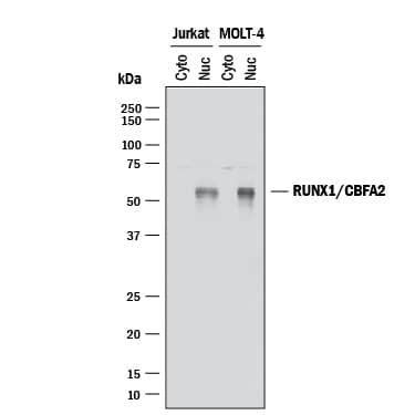
Detection of Human RUNX1/CBFA2 by Western Blot.
Western blot shows cytoplasmic and nuclear extracts from Jurkat human acute T cell leukemia cell line and MOLT-4 human acute lymphoblastic leukemia cell line. PVDF membrane was probed with 2 µg/mL of Goat Anti-Human RUNX1/CBFA2 Antigen Affinity-purified Polyclonal Antibody (Catalog # AF2399) followed by HRP-conjugated Anti-Goat IgG Secondary Antibody (Catalog # HAF017). A specific band was detected for RUNX1/CBFA2 at approximately 55 kDa (as indicated). This experiment was conducted under reducing conditions and using Immunoblot Buffer Group 1.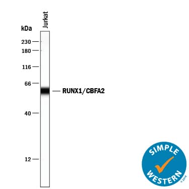
Detection of Human RUNX1/CBFA2 by Simple WesternTM.
Simple Western lane view shows lysates of Jurkat human acute T cell leukemia cell line, loaded at 0.2 mg/mL. A specific band was detected for RUNX1/ CBFA2 at approximately 59 kDa (as indicated) using 20 µg/mL of Goat Anti-Human RUNX1/CBFA2 Antigen Affinity-purified Polyclonal Antibody (Catalog # AF2399) followed by 1:50 dilution of HRP-conjugated Anti-Goat IgG Secondary Antibody (Catalog # HAF109). This experiment was conducted under reducing conditions and using the 12-230 kDa separation system.Applications for Human RUNX1/CBFA2 Antibody
Application
Recommended Usage
Simple Western
20 µg/mL
Sample: Jurkat human acute T cell leukemia cell line
Sample: Jurkat human acute T cell leukemia cell line
Western Blot
2 µg/mL
Sample: Nuclear extracts from Jurkat human acute T cell leukemia cell line and MOLT‑4 human acute lymphoblastic leukemia cell line
Sample: Nuclear extracts from Jurkat human acute T cell leukemia cell line and MOLT‑4 human acute lymphoblastic leukemia cell line
Formulation, Preparation, and Storage
Purification
Antigen Affinity-purified
Reconstitution
Reconstitute at 0.2 mg/mL in sterile PBS. For liquid material, refer to CoA for concentration.
Formulation
Lyophilized from a 0.2 μm filtered solution in PBS with Trehalose. *Small pack size (SP) is supplied either lyophilized or as a 0.2 µm filtered solution in PBS.
Shipping
Lyophilized product is shipped at ambient temperature. Liquid small pack size (-SP) is shipped with polar packs. Upon receipt, store immediately at the temperature recommended below.
Stability & Storage
Use a manual defrost freezer and avoid repeated freeze-thaw cycles.
- 12 months from date of receipt, -20 to -70 °C as supplied.
- 1 month, 2 to 8 °C under sterile conditions after reconstitution.
- 6 months, -20 to -70 °C under sterile conditions after reconstitution.
Background: RUNX1/CBFA2
Long Name
Runt-related Transcription Factor 1
Alternate Names
AML1, CBFA2
Gene Symbol
RUNX1
UniProt
Additional RUNX1/CBFA2 Products
Product Documents for Human RUNX1/CBFA2 Antibody
Product Specific Notices for Human RUNX1/CBFA2 Antibody
For research use only
Loading...
Loading...
Loading...
Loading...
Loading...