
Human/Mouse/Rat Contactin-2/TAG1 Antibody
R&D Systems, part of Bio-Techne | Catalog # AF4439

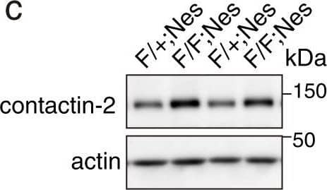
Scientific images may be resized, cropped, or adjusted for brightness or contrast for presentation purposes. These updates are limited to presentation and do not affect the underlying scientific interpretation of the data. Where available, additional source data may be provided upon request.
Conjugate
Catalog #
Key Product Details
Species Reactivity
Validated:
Human, Mouse, Rat
Cited:
Human, Mouse, Rat, Hamster - Mesocricetus auratus (Golden Hamster), Primate, Transgenic Mouse
Applications
Validated:
Immunohistochemistry, Western Blot, Simple Western
Cited:
Immunohistochemistry, Immunohistochemistry-Frozen, Western Blot, Immunocytochemistry, Mass Spectrometry
Label
Unconjugated
Antibody Source
Polyclonal Goat IgG
Product Specifications
Immunogen
Mouse myeloma cell line NS0-derived recombinant mouse Contactin-2/TAG1
Gln31-Ser1014
Accession # Q61330
Gln31-Ser1014
Accession # Q61330
Specificity
Detects mouse and rat Contactin-2 in direct ELISAs. Detects human, mouse, and rat Contactin-2 in Western blots.
Clonality
Polyclonal
Host
Goat
Isotype
IgG
Scientific Data Images for Human/Mouse/Rat Contactin-2/TAG1 Antibody
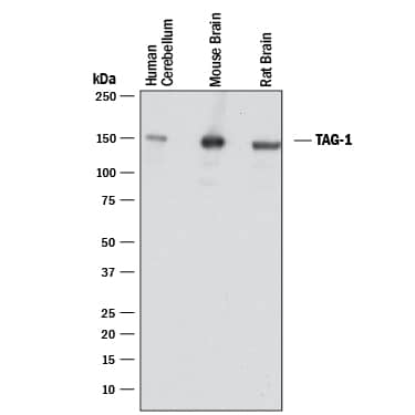
Detection of Human, Mouse, and Rat Contactin‑2/TAG1 by Western Blot.
Western blot shows lysates of human cerebellum tissue, mouse brain tissue, and rat brain tissue. PVDF membrane was probed with 1 µg/mL of Goat Anti-Human/Mouse/Rat Contactin-2/TAG1 Antigen Affinity-purified Polyclonal Antibody (Catalog # AF4439) followed by HRP-conjugated Anti-Goat IgG Secondary Antibody (Catalog # HAF017). A specific band was detected for Contactin-2/TAG1 at approximately 135 kDa (as indicated). This experiment was conducted under reducing conditions and using Immunoblot Buffer Group 1.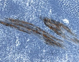
Contactin‑2/TAG1 in Mouse Embryo.
Contactin-2/TAG1 was detected in immersion fixed frozen sections of mouse embryo (E13) using Goat Anti-Human/Mouse/Rat Contactin-2/TAG1 Antigen Affinity-purified Polyclonal Antibody (Catalog # AF4439) at 15 µg/mL overnight at 4 °C. Tissue was stained using the Anti-Goat HRP-DAB Cell & Tissue Staining Kit (brown; Catalog # CTS008) and counterstained with hematoxylin (blue). Specific staining was localized to muscle cells in proximity to ribs. View our protocol for Chromogenic IHC Staining of Frozen Tissue Sections.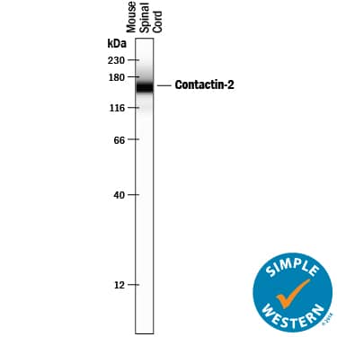
Detection of Mouse Contactin-2/TAG1 by Simple WesternTM.
Simple Western lane view shows lysates of mouse spinal cord tissue, loaded at 0.2 mg/mL. A specific band was detected for Contactin-2/TAG1 at approximately 162 kDa (as indicated) using 10 µg/mL of Goat Anti-Human/Mouse/Rat Contactin-2/TAG1 Antigen Affinity-purified Polyclonal Antibody (Catalog # AF4439) followed by 1:50 dilution of HRP-conjugated Anti-Goat IgG Secondary Antibody (Catalog # HAF109). This experiment was conducted under reducing conditions and using the 12-230 kDa separation system.Applications for Human/Mouse/Rat Contactin-2/TAG1 Antibody
Application
Recommended Usage
Immunohistochemistry
5-15 µg/mL
Sample:
Sample:
Immersion fixed frozen sections of mouse embryo (E13)
Simple Western
10 µg/mL
Sample: Mouse spinal cord tissue
Sample: Mouse spinal cord tissue
Western Blot
1 µg/mL
Sample: Human cerebellum tissue, Mouse brain tissue, and Rat brain tissue
Sample: Human cerebellum tissue, Mouse brain tissue, and Rat brain tissue
Reviewed Applications
Read 2 reviews rated 4.5 using AF4439 in the following applications:
Formulation, Preparation, and Storage
Purification
Antigen Affinity-purified
Reconstitution
Reconstitute at 0.2 mg/mL in sterile PBS. For liquid material, refer to CoA for concentration.
Formulation
Lyophilized from a 0.2 μm filtered solution in PBS with Trehalose. *Small pack size (SP) is supplied either lyophilized or as a 0.2 µm filtered solution in PBS.
Shipping
Lyophilized product is shipped at ambient temperature. Liquid small pack size (-SP) is shipped with polar packs. Upon receipt, store immediately at the temperature recommended below.
Stability & Storage
Use a manual defrost freezer and avoid repeated freeze-thaw cycles.
- 12 months from date of receipt, -20 to -70 °C as supplied.
- 1 month, 2 to 8 °C under sterile conditions after reconstitution.
- 6 months, -20 to -70 °C under sterile conditions after reconstitution.
Background: Contactin-2/TAG1
Contactin-2 (CNTN2), also called TAG1 (transient axonal glycoprotein), TAX1 (transiently-expressed axonal glycoprotein), or axonin-1, is a 135 kDa glycosyl-phosphatidylinositol (GPI)- anchored cell adhesion molecule that belongs to the contactin subfamily within the immunoglobulin (Ig) protein superfamily (1-3). Mouse Contactin-2 cDNA encodes a 30 amino acid (aa) signal peptide, a 984 aa mature secreted protein with 6 Ig-like domains followed by 4 fibronectin type III-like repeats, and a 26 aa C-terminal GPI anchor pro-sequence. GPI-specific phospholipase activity can release soluble, active Contactin-2 from the membrane (2). Mature mouse Contactin-2 shares approximately 93%, 97%, and 77% aa sequence identity with human, rat, and chicken Contactin-2, respectively. During development, Contactin-2 is expressed by a subset of neuronal populations in the central nervous system (CNS) and peripheral nervous system (PNS), particularly during initial phases of axon outgrowth (3-5). Both the 135 kDa form and a 90 kDa form are also upregulated in response to CNS injury in the adult (6). Data support a role for Contactin-2 in axon pathfinding, neurite outgrowth and adhesion, especially in the CNS (3-6). In mature myelinated fibers, Contactin-2 is expressed by oligodendrocytes and Schwann cells, which are myelinating glial cells of the CNS and PNS, respectively (7, 8). It is enriched in the juxtaparanodal regions, where it recruits contactin-associated protein 2 (caspr2), a transmembrane neurexin involved in cell adhesion and intercellular communication (7-10). The axonal Contactin-2 interacts in cis with caspr2 and in trans with another Contactin-2 on the glial membrane (8). This ternary complex is required for the accumulation and organization of K+ channels in the juxtaparanodes (9).
References
- Wolfer, D. and R.J. Giger (1994) Swissprot Accession # Q61330.
- Hasler, T.H. et al. (1993) Eur. J. Biochem. 211:329.
- Karagogeos, D. (2003) Front. Biosci. 8:s1304.
- Liu, Y. and M.C. Halloran (2005) J. Neurosci. 25:10556.
- Denaxa, M. et al. (2005) Dev. Biol. 288:87.
- Soares, S. et al. (2005) Eur. J. Neurosci. 21:1169.
-
Traka, M. et al. (2002) J. Neurosci. 22:3016.
-
Poliak, S. and E. Peles (2003) Nat. Rev. Neurosci. 4:968.
-
Traka, M. et al. (2003) J. Cell Biol. 162:1161.
-
Poliak, S. et al. (2003) J. Cell Biol. 162:1149.
Alternate Names
Axonin-1, AXT, CNTN2, Contactin2, TAG1, TAX1
Gene Symbol
CNTN2
UniProt
Additional Contactin-2/TAG1 Products
Product Documents for Human/Mouse/Rat Contactin-2/TAG1 Antibody
Product Specific Notices for Human/Mouse/Rat Contactin-2/TAG1 Antibody
For research use only
Loading...
Loading...
Loading...
Loading...
Loading...