
Human MMP-1 Antibody
R&D Systems, part of Bio-Techne | Catalog # AF901



Scientific images may be resized, cropped, or adjusted for brightness or contrast for presentation purposes. These updates are limited to presentation and do not affect the underlying scientific interpretation of the data. Where available, additional source data may be provided upon request.
Conjugate
Catalog #
Key Product Details
Validated by
Knockout/Knockdown
Species Reactivity
Validated:
Human
Cited:
Human
Applications
Validated:
Knockout Validated, Immunohistochemistry, Western Blot, ELISA Capture (Matched Antibody Pair), Simple Western
Cited:
Immunohistochemistry, Western Blot, ELISA Development, ELISA Microarray Development
Label
Unconjugated
Antibody Source
Polyclonal Goat IgG
Product Specifications
Immunogen
Mouse myeloma cell line NS0-derived recombinant human MMP-1
Phe20-Asn469
Accession # P03956
Phe20-Asn469
Accession # P03956
Specificity
Detects human MMP-1 in ELISAs and Western blots. In sandwich immunoassays, less than 0.5% cross-reactivity with recombinant human (rh) MMP‑2, rhMMP-3, rhMMP-7, rhMMP-8, rhMMP-9, rhMMP-10, rhMMP-12, rhMMP-13, and rhMMP-16 is observed.
Clonality
Polyclonal
Host
Goat
Isotype
IgG
Scientific Data Images for Human MMP-1 Antibody
Detection of Human MMP‑1 by Western Blot.
Western blot shows lysates of PC-3 human prostate cancer cell line and HEK001 human epidermal keratinocyte cell line. PVDF membrane was probed with 1 µg/mL of Goat Anti-Human MMP-1 Antigen Affinity-purified Polyclonal Antibody (Catalog # AF901) followed by HRP-conjugated Anti-Goat IgG Secondary Antibody (Catalog # HAF017). A specific band was detected for MMP-1 at approximately 50 kDa (as indicated). This experiment was conducted under reducing conditions and using Immunoblot Buffer Group 1.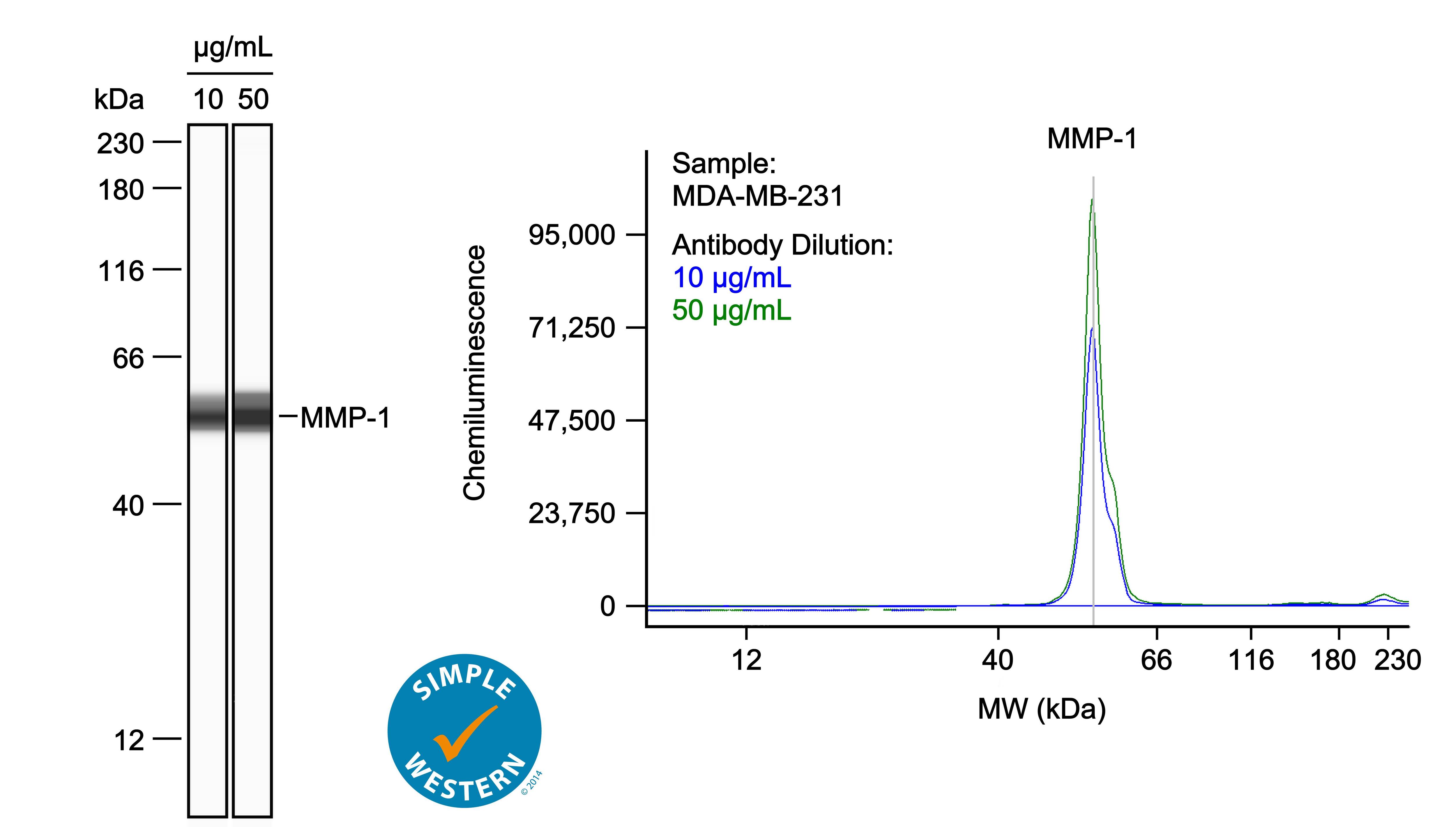
Detection of Human MMP-1 by Simple WesternTM.
Left: Simple Western lane view shows lysates of MDA-MB-231 human breast cancer cell line, loaded at 0.1 mg/ml. A specific band was detected for MMP-1 at approximately 55 kDa (as indicated) using both 10 µg/ml and 50 µg/ml of Goat Anti-Human MMP-1 Antigen Affinity-purified Polyclonal Antibody (Catalog # AF901) followed by HRP-conjugated Donkey Anti-Goat Secondary Antibody (Catalog # 043-522-2). This experiment was conducted under reducing conditions and using the 12-230kDa separation system. Right: Simple Western electropherogram showing the same Goat Anti-Human MMP-1 Antigen Affinity-purified Polyclonal Antibody (Catalog # AF901) tested at 10 µg/ml (blue line) and 50 µg/ml (green line) in the MDA-MB-231 human breast cancer cell line.
MMP‑1 in Human Ovarian Cancer Tissue.
MMP-1 was detected in immersion fixed paraffin-embedded sections of human ovarian cancer tissue using 15 µg/mL Goat Anti-Human MMP-1 Antigen Affinity-purified Polyclonal Antibody (Catalog # AF901) overnight at 4 °C. Tissue was stained with the Anti-Goat HRP-DAB Cell & Tissue Staining Kit (brown; Catalog # CTS008) and counterstained with hematoxylin (blue). View our protocol for Chromogenic IHC Staining of Paraffin-embedded Tissue Sections.Applications for Human MMP-1 Antibody
Application
Recommended Usage
Immunohistochemistry
5-15 µg/mL
Sample: Immersion fixed paraffin-embedded sections of human ovarian cancer tissue and immersion fixed paraffin-embedded sections of human prostate
Sample: Immersion fixed paraffin-embedded sections of human ovarian cancer tissue and immersion fixed paraffin-embedded sections of human prostate
Knockout Validated
MMP-1 is specifically detected in PC‑3 human prostate cancer parental cell line but is not detectable in MMP-1 knockout PC‑3 cell line.
Simple Western
10-50 µg/mL
Sample: MDA-MB-231 human breast cancer cell line
Sample: MDA-MB-231 human breast cancer cell line
Western Blot
1 µg/mL
Sample: PC‑3 human prostate cancer cell line and HEK001 human epidermal keratinocyte cell line
Sample: PC‑3 human prostate cancer cell line and HEK001 human epidermal keratinocyte cell line
Human MMP-1 Sandwich Immunoassay
Please Note: Optimal dilutions of this antibody should be experimentally determined.
Reviewed Applications
Read 2 reviews rated 4 using AF901 in the following applications:
Formulation, Preparation, and Storage
Purification
Antigen Affinity-purified
Reconstitution
Reconstitute at 0.2 mg/mL in sterile PBS. For liquid material, refer to CoA for concentration.
Formulation
Lyophilized from a 0.2 μm filtered solution in PBS with Trehalose. See Certificate of Analysis for details.
*Small pack size (-SP) is supplied either lyophilized or as a 0.2 µm filtered solution in PBS.
*Small pack size (-SP) is supplied either lyophilized or as a 0.2 µm filtered solution in PBS.
Shipping
Lyophilized product is shipped at ambient temperature. Liquid small pack size (-SP) is shipped with polar packs. Upon receipt, store immediately at the temperature recommended below.
Stability & Storage
Use a manual defrost freezer and avoid repeated freeze-thaw cycles.
- 12 months from date of receipt, -20 to -70 °C as supplied.
- 1 month, 2 to 8 °C under sterile conditions after reconstitution.
- 6 months, -20 to -70 °C under sterile conditions after reconstitution.
Background: MMP-1
Matrix metalloproteinases are a family of zinc and calcium dependent endopeptidases with the combined ability to degrade all the components of the extracellular matrix. MMP-1 (interstitial collagenase), can degrade a broad range of substrates including types I, II, III, VII, VIII, and X collagens as well as casein, gelatin,
alpha-1 antitrypsin, myelin basic protein, L-Selectin, pro-TNF, IL-1 beta, IGFBP-3, IGFBP-5, pro-MMP-2, and pro-MMP-9. A significant role of MMP-1 is the degradation of fibrillar collagens in extracellular matrix remodeling, characterized by the cleavage of the interstitial collagen triple helix into ¾, ¼ fragments. However, as the list of substrates above illustrates, the role of MMP-1 is more diverse than originally envisaged, and may involve enzyme cascades, cytokine regulation, and cell surface molecule modulation. MMP-1 is expressed by fibroblasts, keratinocytes, endothelial cells, monocytes, and macrophages. Structurally, MMP-1 may be divided into several distinct domains; a pro-domain which is cleaved upon activation; a catalytic domain containing the zinc binding site; a short hinge region and a carboxyl terminal (hemopexin-like) domain.
References
- Cawston, T.E. (2004) in Interstitial Collagenase. Barrett, A.J. et al. (eds): Handbook of Proteolytic Enzymes, San Diego: Academic Press, p. 472.
Long Name
Matrix Metalloproteinase 1
Alternate Names
MMP1
Gene Symbol
MMP1
UniProt
Additional MMP-1 Products
Product Documents for Human MMP-1 Antibody
Product Specific Notices for Human MMP-1 Antibody
For research use only
Loading...
Loading...
Loading...
Loading...
Loading...