
Human MICA/B Antibody
R&D Systems, part of Bio-Techne | Catalog # MAB13001


Scientific images may be resized, cropped, or adjusted for brightness or contrast for presentation purposes. These updates are limited to presentation and do not affect the underlying scientific interpretation of the data. Where available, additional source data may be provided upon request.
Conjugate
Catalog #
Key Product Details
Species Reactivity
Validated:
Human
Cited:
Human
Applications
Validated:
Immunohistochemistry, Blockade of Receptor-ligand Interaction, Flow Cytometry, CyTOF-ready
Cited:
Immunohistochemistry, Western Blot, Neutralization, Flow Cytometry, Immunocytochemistry, Immunoprecipitation
Label
Unconjugated
Antibody Source
Monoclonal Mouse IgG2A Clone # 159207
Product Specifications
Immunogen
BaF3 mouse pro-B cell line transfected with human MICA
Accession # Q29983
Accession # Q29983
Specificity
Detects human MICA/B in direct ELISAs.
Clonality
Monoclonal
Host
Mouse
Isotype
IgG2A
Endotoxin Level
<0.10 EU per 1 μg of the antibody by the LAL method.
Scientific Data Images for Human MICA/B Antibody
Detection of MICA/B in K562 Human Cell Line by Flow Cytometry.
K562 human chronic myelogenous leukemia cell line was stained with Mouse Anti-Human MICA/B Monoclonal Antibody (Catalog # MAB13001, filled histogram) or isotype control antibody (Catalog # MAB003, open histogram), followed by Phycoerythrin-conjugated Anti-Mouse IgG Secondary Antibody (Catalog # F0102B). View our protocol for Staining Membrane-associated Proteins.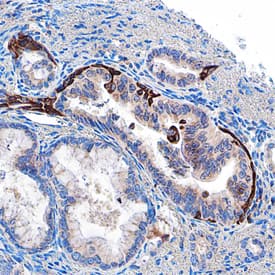
MICA/B in Human Pancreas.
MICA/B was detected in immersion fixed paraffin-embedded sections of human pancreas using Mouse Anti-Human MICA/B Monoclonal Antibody (Catalog # MAB13001) at 15 µg/mL overnight at 4 °C. Before incubation with the primary antibody, tissue was subjected to heat-induced epitope retrieval using Antigen Retrieval Reagent-Basic (Catalog # CTS013). Tissue was stained using the Anti-Mouse HRP-DAB Cell & Tissue Staining Kit (brown; Catalog # CTS002) and counterstained with hematoxylin (blue). Specific staining was localized to exocrine cells. View our protocol for Chromogenic IHC Staining of Paraffin-embedded Tissue Sections.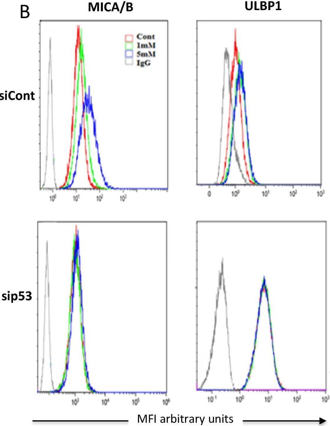
Detection of MICA/B by Flow Cytometry
Decreasing p53 levels prevents DCA-induced stress ligands upregulation. (A) OCI-AML3 cells were transfected with siRNA for p53 or control siRNA and 24 h later were treated with 1and 5 mM DCA for 3 days. Protein and mRNA levels were quantified as described in Supplemental Fig. 1 and Fig. 1, respectively. Bar graphs denote the % of mRNA compared to non-treated cells and represent means ± SD of 3 independent experiments; *p < 0.05, **p < 0.01, ***p < 0.005 student t-test compare to non-treated cells. (B) The expression of stress ligands in cell membrane were analyzed by FACs. Image collected and cropped by CiteAb from the following open publication (https://www.nature.com/articles/s41598-022-07281-6), licensed under a CC-BY license. Not internally tested by R&D Systems.Applications for Human MICA/B Antibody
Application
Recommended Usage
Blockade of Receptor-ligand Interaction
In a functional ELISA, 0.03-0.1 µg/mL of this antibody will block 50% of the binding of 50 ng/mL of Recombinant Biotinylated Human MICA Fc Chimera to immobilized Recombinant Human NKG2D Fc Chimera (Catalog # 1299‑NK) coated at 2 µg/mL (100 µL/well). At 0.4 μg/mL, this antibody will block >90% of the binding.
CyTOF-ready
Ready to be labeled using established conjugation methods. No BSA or other carrier proteins that could interfere with conjugation.
Flow Cytometry
0.25 µg/106 cells
Sample: K562 human chronic myelogenous leukemia cell line
Sample: K562 human chronic myelogenous leukemia cell line
Immunohistochemistry
8-25 µg/mL
Sample: Immersion fixed paraffin-embedded sections of human pancreas
Sample: Immersion fixed paraffin-embedded sections of human pancreas
Formulation, Preparation, and Storage
Purification
Protein A or G purified from hybridoma culture supernatant
Reconstitution
Reconstitute at 0.5 mg/mL in sterile PBS. For liquid material, refer to CoA for concentration.
Formulation
Lyophilized from a 0.2 μm filtered solution in PBS with Trehalose. *Small pack size (SP) is supplied either lyophilized or as a 0.2 µm filtered solution in PBS.
Shipping
Lyophilized product is shipped at ambient temperature. Liquid small pack size (-SP) is shipped with polar packs. Upon receipt, store immediately at the temperature recommended below.
Stability & Storage
Use a manual defrost freezer and avoid repeated freeze-thaw cycles.
- 12 months from date of receipt, -20 to -70 °C as supplied.
- 1 month, 2 to 8 °C under sterile conditions after reconstitution.
- 6 months, -20 to -70 °C under sterile conditions after reconstitution.
Background: MICA/B
Long Name
MHC Class I-related Protein A and B
Alternate Names
MHC class I chain-related protein A, MHC class I chain-related protein B, MIC, MICA, MICB, PERB11, PERB11.1, PERB11.2
Entrez Gene IDs
4276 (Human)
Gene Symbol
MICA
UniProt
Additional MICA/B Products
Product Documents for Human MICA/B Antibody
Product Specific Notices for Human MICA/B Antibody
For research use only
Loading...
Loading...
Loading...
Loading...
Loading...
Loading...