
Human IGF-II R/IGF2R Fluorescein-conjugated Antibody
R&D Systems, part of Bio-Techne | Catalog # FAB2447F

Conjugate
Catalog #
Key Product Details
Species Reactivity
Validated:
Human
Cited:
Human, Mouse
Applications
Validated:
Flow Cytometry
Cited:
Flow Cytometry
Label
FITC (Excitation = 488 nm, Emission = 515-545 nm)
Antibody Source
Polyclonal Goat IgG
Product Specifications
Immunogen
Mouse myeloma cell line NS0-derived recombinant human IGF-II R
Ser1510-Phe2108
Accession # P11717
Ser1510-Phe2108
Accession # P11717
Specificity
Detects human IGF-II R in direct ELISAs and Western blots.
Clonality
Polyclonal
Host
Goat
Isotype
IgG
Scientific Data Images for Human IGF-II R/IGF2R Fluorescein-conjugated Antibody
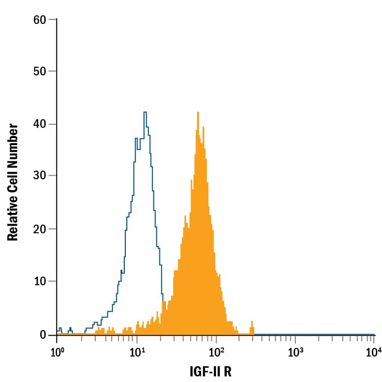
Detection of IGF‑II R in Human Blood Monocytes by Flow Cytometry.
Human peripheral blood monocytes were stained with Goat Anti-Human IGF-II R Fluorescein-conjugated Antigen Affinity-purified Polyclonal Antibody (Catalog # FAB2447F, filled histogram) or isotype control antibody (Catalog # IC108F, open histogram). View our protocol for Staining Membrane-associated Proteins.Applications for Human IGF-II R/IGF2R Fluorescein-conjugated Antibody
Application
Recommended Usage
Flow Cytometry
10 µL/106 cells
Sample: Human peripheral blood monocytes
Sample: Human peripheral blood monocytes
Spectra Viewer
Plan Your Experiments
Use our spectra viewer to interactively plan your experiments, assessing multiplexing options. View the excitation and emission spectra for our fluorescent dye range and other commonly used dyes.
Spectra Viewer
Formulation, Preparation, and Storage
Purification
Antigen Affinity-purified
Formulation
Supplied in a saline solution containing BSA and Sodium Azide.
Shipping
The product is shipped with polar packs. Upon receipt, store it immediately at the temperature recommended below.
Stability & Storage
Protect from light. Do not freeze.
- 12 months from date of receipt, 2 to 8 °C as supplied.
Background: IGF-II R/IGF2R
The type 2 Insulin-like Growth Factor Receptor (IGF-II R; also known as cation-independent mannose-6 phosphate receptor/CI-MPR) is a 300 kDa member of the P-type lectin family of molecules. P-type lectins generate functional eukaryotic lysosomes by binding and sorting lysosomal enzymes expressing phosphorylated mannose residues (M6P) (1-3). IGF-II R is a type I transmembrane glycoprotein that contains a 2,264 amino acid (aa) extracellular region, a 23 aa transmembrane segment and a 124 aa cytoplasmic tail (4, 5). The extracellular region consists of 15 contiguous “binding” repeats of about 150 aa each. The odd-numbered repeats interact with “ligands” while the even-numbered repeats likely generate a nondisulfide homodimer in the membrane (1). Repeat #11 binds IGF‑II, while repeats 3 and 9 bind mannose-6 phosphate; repeat #13 contains a fibronectin type II motif and assists in IGF-II binding (1, 2). In the extracellular region of IGF-II R expressed by R&D Systems (600 amino acids), human IGF-II R is 85% identical to both mouse and bovine IGF-II R. This expressed region includes binding repeats #11, 12, and 13. In addition to IGF-II, CI-MPR/IGF-II R binds non-M6P containing ligands such as retinoic acid, urokinase-type plasminogen-activator receptor and plasminogen, plus M6P-containing molecules such as lysosomal enzymes, TGF-beta 1 precursor, proliferin, LIF, CD26, herpes simplex glycoprotein D, and granzymes A and B (2, 6). IGF-II R regulates many diverse biological functions that range from intracellular trafficking to the internalization of extracellular factors and modulation of cellular responses. It delivers newly synthesized M6P-tagged lysosomal enzymes from the trans-golgi network to endosomes, and facilitates the clearance of extracellular lysosomal and matrix degrading enzymes by internalization into clathrin-coated vesicles and delivery into endosomes. With respect to IGF-II biology, it would appear that IGF-II R is principally a regulator of local IGF-II levels, targeting IGF-II for destruction in lysosomes (2). However, some evidence suggests the receptor will signal via G-proteins, an effect that has yet to be conclusively shown (6).
References
- Ghosh, P. et al. (2003) Nat. Rev. Mol. Cell. Biol. 4:202.
- Dahms, N.M. and M.K. Hancock (2002) Biochim. Biophys. Acta. 1572:317.
- Zaina, S. and J. Nilsson (2003) Curr. Opin. Lipidol. 14:483.
- Morgan, D.O. et al. (1987) Nature 329:301.
- Oshima, A. et al. (1988) J. Biol. Chem. 263:2553.
- Hawkes, C. and S. Kar (2004) Brain Res. Rev. 44:117.
Long Name
Insulin-like Growth Factor II Receptor
Alternate Names
CD222, CIMPR, IGF-IIR, IGF2R, IGFIIR, M6P-R, MPRI
Entrez Gene IDs
3482 (Human)
Gene Symbol
IGF2R
UniProt
Additional IGF-II R/IGF2R Products
Product Documents for Human IGF-II R/IGF2R Fluorescein-conjugated Antibody
Product Specific Notices for Human IGF-II R/IGF2R Fluorescein-conjugated Antibody
For research use only
Loading...
Loading...
Loading...
Loading...
Loading...