Human 14-3-3 zeta Antibody
R&D Systems, part of Bio-Techne | Catalog # MAB2669


Conjugate
Catalog #
Key Product Details
Species Reactivity
Human
Applications
Immunohistochemistry, Western Blot, Simple Western
Label
Unconjugated
Antibody Source
Monoclonal Mouse IgG2B Clone # 818515
Product Specifications
Immunogen
E. coli-derived recombinant human 14-3-3 zeta
Asp2-Asn245
Accession # P63104
Asp2-Asn245
Accession # P63104
Specificity
Detects human 14-3-3 zeta in direct ELISAs and Western blots. Detects Mouse and Rat 14-3-3 zeta in Western Blots. In direct ELISAs, nocross-reactivity with recombinant human 14-3-3 beta, theta, eta, gamma, sigma, or epsilon is observed.
Clonality
Monoclonal
Host
Mouse
Isotype
IgG2B
Scientific Data Images for Human 14-3-3 zeta Antibody
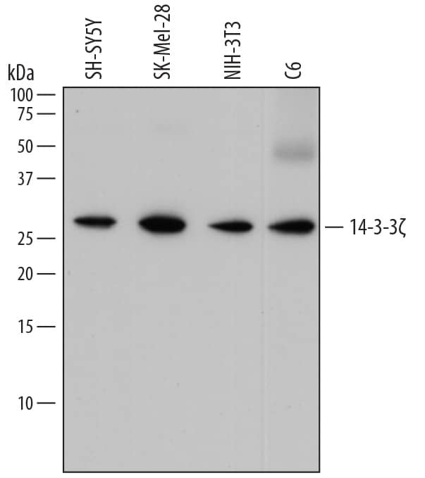
Detection of Human, Mouse, and Rat 14‑3‑3 zeta by Western Blot.
Western blot shows lysates of SH-SY5Y human neuroblastoma cell line, SK-Mel-28 human malignant melanoma cell line, NIH-3T3 mouse embryonic fibroblast cell line, and C6 rat glioma cell line. PVDF membrane was probed with 0.2 µg/mL of Mouse Anti-Human 14-3-3 zeta Monoclonal Antibody (Catalog # MAB2669) followed by HRP-conjugated Anti-Mouse IgG Secondary Antibody (Catalog # HAF018). A specific band was detected for 14-3-3 zeta at approximately 27 kDa (as indicated). This experiment was conducted under reducing conditions and using Immunoblot Buffer Group 1.14‑3‑3 zeta in Human Squamous Cell Carcinoma.
14-3-3 zeta was detected in immersion fixed paraffin-embedded sections of human squamous cell carcinoma using Mouse Anti-Human 14-3-3 zeta Monoclonal Antibody (Catalog # MAB2669) at 15 µg/mL overnight at 4 °C. Before incubation with the primary antibody, tissue was subjected to heat-induced epitope retrieval using Antigen Retrieval Reagent-Basic (Catalog # CTS013). Tissue was stained using the Anti-Mouse HRP-DAB Cell & Tissue Staining Kit (brown; Catalog # CTS002) and counter-stained with hematoxylin (blue). Specific staining was localized to nuclei and plasma membrane. View our protocol for Chromogenic IHC Staining of Paraffin-embedded Tissue Sections.Detection of Human 14‑3‑3 zeta by Simple WesternTM.
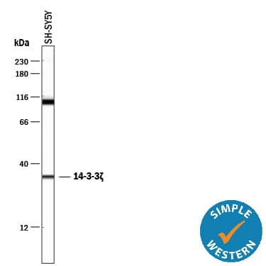
Simple Western lane view shows lysates of SH‑SY5Y human neuroblastoma cell line, loaded at 0.5 mg/mL. A specific band was detected for 14‑3‑3 zeta at approximately 34 kDa (as indicated) using 2 µg/mL of Mouse Anti-Human 14‑3‑3 zeta Monoclonal Antibody (Catalog # MAB2669). This experiment was conducted under reducing conditions and using the 12-230 kDa separation system.Applications for Human 14-3-3 zeta Antibody
Application
Recommended Usage
Immunohistochemistry
8-25 µg/mL
Sample: Immersion fixed paraffin-embedded sections of human squamous cell carcinoma
Sample: Immersion fixed paraffin-embedded sections of human squamous cell carcinoma
Simple Western
2 µg/mL
Sample: SH‑SY5Y human neuroblastoma cell line
Sample: SH‑SY5Y human neuroblastoma cell line
Western Blot
0.2 µg/mL
Sample: SH‑SY5Y human neuroblastoma cell line, SK‑Mel‑28 human malignant melanoma cell line, NIH‑3T3 mouse embryonic fibroblast cell line, and C6 rat glioma cell line
Sample: SH‑SY5Y human neuroblastoma cell line, SK‑Mel‑28 human malignant melanoma cell line, NIH‑3T3 mouse embryonic fibroblast cell line, and C6 rat glioma cell line
Reviewed Applications
Read 2 reviews rated 5 using MAB2669 in the following applications:
Formulation, Preparation, and Storage
Purification
Protein A or G purified from hybridoma culture supernatant
Reconstitution
Sterile PBS to a final concentration of 0.5 mg/mL. For liquid material, refer to CoA for concentration.
Formulation
Lyophilized from a 0.2 μm filtered solution in PBS with Trehalose. *Small pack size (SP) is supplied either lyophilized or as a 0.2 µm filtered solution in PBS.
Shipping
Lyophilized product is shipped at ambient temperature. Liquid small pack size (-SP) is shipped with polar packs. Upon receipt, store immediately at the temperature recommended below.
Stability & Storage
Use a manual defrost freezer and avoid repeated freeze-thaw cycles.
- 12 months from date of receipt, -20 to -70 °C as supplied.
- 1 month, 2 to 8 °C under sterile conditions after reconstitution.
- 6 months, -20 to -70 °C under sterile conditions after reconstitution.
Background: 14-3-3 zeta
Additional 14-3-3 zeta Products
Product Documents for Human 14-3-3 zeta Antibody
Product Specific Notices for Human 14-3-3 zeta Antibody
For research use only
Loading...
Loading...
Loading...
Loading...

