
GloLIVE Human Pluripotent Stem Cell Live Cell Imaging Kit
R&D Systems, part of Bio-Techne | Catalog # SC023B


Discontinued Product
SC023B has been discontinued.
View all Pluripotent Stem Cells products.
Key Product Details
Species
Human
Product Summary for GloLIVE Human Pluripotent Stem Cell Live Cell Imaging Kit
Kit Summary
To verify pluripotency in live, unfixed human stem cells and continue in culture.
Key Benefits
- Verify human stem cell pluripotency in 30 minutes
- Includes 4 pluripotent stem cell-specific fluorochrome-conjugated antibodies
- Single-step protocol
- No adverse effects on stemness or proliferation
- Pick pluripotent human stem cell colonies with increased confidence

Why Analyze Human Stem Cell Pluripotency by Live Cell Staining Using Stem Cell Markers?
Maintaining pluripotent human embryonic stem (ES) cells and deriving induced pluripotent stem (iPS) cells requires colony selection and cell expansion. Manually picking colonies that contain pluripotent stem cells can be achieved by analyzing colony morphology. However, colony morphology is not truly indicative of pluripotency, since cells can naturally differentiate within the colony. Moreover, colony picking is a time-consuming and labor-intensive process.
Designed to verify pluripotency in live cells, the GloLIVE™ Human Pluripotent Stem Cell Live Cell Imaging Kit increases research efficiency and success by allowing single-step staining of unfixed cells.
Live pluripotent stem cell imaging:
- Promotes the selection of high quality, undifferentiated stem cell colonies to reduce experimental variation.
- Verifies stem cell pluripotency in 30 minutes, ensuring efficient use of time and reagents.
- Provides increased confidence in pluripotent status through the use of positive and negative markers.
- Allows for the continued culture of pluripotent cells with no adverse effects on cell proliferation or stemness following staining.
Kit Contents
This kit contains the following azide-free, fluorochrome-conjugated antibodies to verify human stem cell pluripotency:
Positive Markers
- NL493-conjugated Mouse Anti-Human SSEA-4
- NL557-conjugated Mouse Anti-Human TRA-1-60(R)
- NL493-conjugated Mouse Anti-Human TRA-1-81
Negative Marker
- NL557-conjugated Mouse Anti-Human SSEA-1
Each antibody is supplied as a 50X stock; enough for 25 assays when used in 500 µL staining volume per assay. Each 1X antibody solution has an endotoxin level of < 5 EU/mL.
Stability and Storage
Reagents are stable for 12 months from date of receipt when stored in the dark at 2° C to 8°.
Data Examples
 View Larger Image |
Human Induced Pluripotent Stem Cells Express TRA-1-81. |
 View Larger Image |
Verification of Pluripotency in Human Induced Pluripotent Stem Cells. |
 View Larger Image |
Analysis of Human Embryonic Stem Cell Growth and Stemness Post-staining. |
Scientific Data Images for GloLIVE Human Pluripotent Stem Cell Live Cell Imaging Kit
Human Induced Pluripotent Stem Cells Express TRA-1-81.
JOY1 human induced pluripotent stem cells were stained with the NL493-conjugated TRA-1-81 (green) antibody included in the GloLIVE™Human Induced Pluripotent Stem Cell Live Cell Imaging Kit. Cells were counterstained with Hoechst 33342 (blue).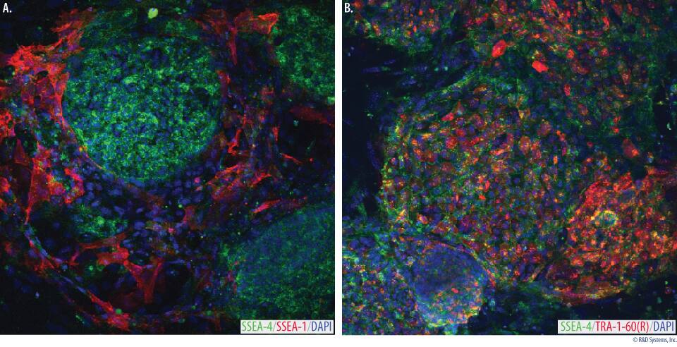
Verification of Pluripotency in Human Induced Pluripotent Stem Cells.
iPS2 human induced pluripotent stem cells were grown on irradiated mouse embryonic fibroblasts (Catalog # PSC001), and stained using antibodies included in the Human Pluripotent Stem Cell Live Cell Imaging Kit.A.iPS2 cells stained with the NL493-conjugated SSEA-4 (green) and NL557-conjugated SSEA-1 (red) antibodies.B.iPS2 cells stained with the NL493-conjugated SSEA-4 (green) and NL557-conjugated TRA-1-60(R) (red) antibodies. The cells were counterstained with Hoechst 33342 (blue). The colonies are positive for the stem cell markers SSEA-4 and TRA-1-60(R) and are negative for SSEA-1, suggesting that these colonies primarily contain undifferentiated human stem cells.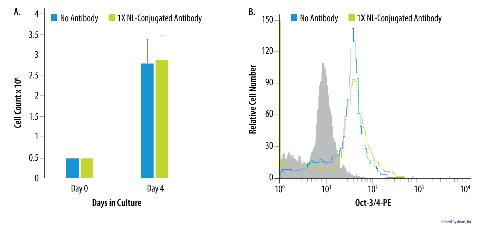
Analysis of Human Embryonic Stem Cell Growth and Stemness Post-staining.
BG01V human embryonic stem cells were plated in triplicate in 6 well plates at 0.5 x 10⁶ cells per well on Day 0. On Day 1, cells were stained with (green bars) or without (blue bars) 1X NL-conjugated antibody. On Day 4, cells were harvested and counted using a hemocytometer. There was no effect of SSEA-4, TRA-1-60(R), or SSEA-1 staining on cell proliferation. Error bars indicate standard deviation.B.BG01V human embryonic stem cells were stained with (green histogram) or without (blue histogram) 1X NL-conjugated antibody and cultured for 3 days. Cells were then harvested and stained using the pluripotent marker PE-conjugated Rat Anti-Human/Mouse Oct-3/4 Monoclonal Antibody (Catalog # IC1759P; open histogram) or a Rat IgG2B PE-conjugated Isotype Control (Catalog # IC013P; filled histogram). Staining with SSEA-4, TRA-1-60(R), or SSEA-1 had no effect on stemness as analyzed by Oct-3/4 expression. Stemness post-staining was also verified by SSEA-4 and SSEA-1 expression (data not shown).Formulation, Preparation, and Storage
Shipping
The product is shipped with polar packs. Upon receipt, store it immediately at the temperature recommended below.
Storage
Store the unopened product at 2 - 8 °C. Do not use past expiration date.
Background: Pluripotent Stem Cells
Embryonic stem (ES) cells are pluripotent stem cells derived from the inner cell mass of pre-implantation embryos. Induced pluripotent stem (iPS) cells can be generated by somatic cell reprogramming following the exogenous expression of specific transcription factors (Oct-3/4, KLF4, SOX2, and c-Myc). These cell types are capable of unlimited, undifferentiated proliferation in vitro and still maintain the capacity to differentiate into a wide variety of somatic cells. In this capacity, pluripotent stem cells have widespread clinical potential for the treatments of heart disease, diabetes, spinal cord injury, and a variety of neurodegenerative disorders.
R&D Systems offers a wide range of products to support pluripotent stem cell culture and differentiation. Mouse embryonic fibroblasts may be used to maintain and expand pluripotent stem cells in an undifferentiated state. We also offer defined culture media, which are specifically optimized for use with human or rodent pluripotent stem cells. In addition, R&D Systems offers a variety of products to assess differentiation status and identify specific stem cell types of interest, including panels of marker antibodies, primer pairs, multi-color flow cytometry kits, and specialized verification kits.
Additional Pluripotent Stem Cells Products
Product Documents for GloLIVE Human Pluripotent Stem Cell Live Cell Imaging Kit
Product Specific Notices for GloLIVE Human Pluripotent Stem Cell Live Cell Imaging Kit
For research use only
Loading...
Loading...
Loading...
Loading...
Loading...