Donkey anti-Rabbit IgG (H+L) Secondary Antibody
Novus Biologicals, part of Bio-Techne | Catalog # NBP1-72696
Conjugate
Catalog #
Key Product Details
Species Reactivity
Rabbit
Applications
Dot Blot, ELISA, Immunohistochemistry, Western Blot
Label
Unconjugated
Antibody Source
Polyclonal Donkey IgG
Concentration
Please see the vial label for concentration. If unlisted please contact technical services.
Product Specifications
Immunogen
Rabbit IgG whole molecule
Clonality
Polyclonal
Host
Donkey
Isotype
IgG
Description
This product was prepared from monospecific antiserum by immunoaffinity chromatography using Rabbit IgG coupled to agarose beads followed by solid phase adsorption(s) to remove any unwanted reactivities. Assay by immunoelectrophoresis resulted in a single precipitin arc against anti-Donkey Serum, Rabbit IgG and Rabbit Serum.
Store vial at 4C prior to opening. This product is stable for several weeks at 4C as an undiluted liquid. Dilute only prior to immediate use. For extended storage aliquot contents and freeze at -20C or below. Avoid cycles of freezing and thawing.
Store vial at 4C prior to opening. This product is stable for several weeks at 4C as an undiluted liquid. Dilute only prior to immediate use. For extended storage aliquot contents and freeze at -20C or below. Avoid cycles of freezing and thawing.
Scientific Data Images for Donkey anti-Rabbit IgG (H+L) Secondary Antibody
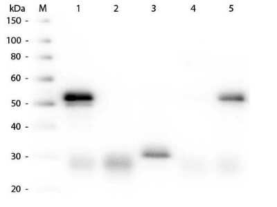
Western Blot: Donkey anti-Rabbit IgG (H+L) Secondary Antibody [Unconjugated] [NBP1-72696] - Lane 1: Rabbit IgG whole molecule. Lane 2: Rabbit IgG F(ab) Fragment. Lane 3: Rabbit IgG F(c) Fragment. Lane 4: Rabbit IgM Whole Molecule. Lane 5: Normal Rabbit Serum. All samples were reduced. Load: 50 ng per lane. Block: incubated with blocking buffer for 30 min at RT. Primary Antibody: Anti-Rabbit IgG (H&L) (DONKEY) Antibody Peroxidase Conjugated 1:5,000 for 60 min at RT. Secondary antibody: None. Predicted/Observed Size: 25 and 50 kDa for Rabbit IgG and Serum, 25 kDa for F(c) and F(ab), 70 and 23 kDa for IgM. Rabbit F(c) migrates slightly higher.
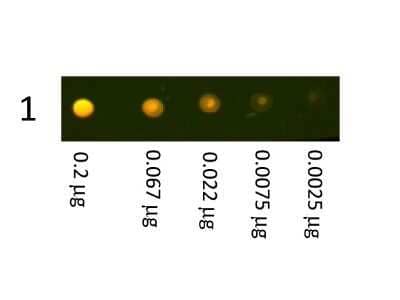
Dot Blot: Donkey anti-Rabbit IgG (H+L) Secondary Antibody [Unconjugated] [NBP1-72696] - Dot Blot showing the detection of Rabbit IgG. A three-fold serial dilution of Rabbit IgG starting at 200 ng was spotted onto 0.45 um nitrocellulose. After blocking in 5% Blotto 1 Hour at 20C, Donkey anti-Rabbit IgG (H+L) Secondary Antibody [Texas Red] was incubated in blocking buffer for Fluorescent Western Blotting and imaged using the Bio-Rad VersaDoc 4000 MP. Image from the Texas Red version of this antibody.
Applications for Donkey anti-Rabbit IgG (H+L) Secondary Antibody
Application
Recommended Usage
ELISA
1:40000 - 1:160000
Immunohistochemistry
1:1000 - 1:5000
Western Blot
1:2000 - 1:10000
Application Notes
This product has been tested by ELISA and is suitable for ELISA, western blot, and immunohistochemistry, as well as other assays requiring lot-to-lot consistency.
Formulation, Preparation, and Storage
Purification
Multi-step
Formulation
0.02 M Potassium Phosphate, 0.15 M Sodium Chloride, pH 7.2
Preservative
0.01% Sodium Azide
Concentration
Please see the vial label for concentration. If unlisted please contact technical services.
Shipping
The product is shipped with polar packs. Upon receipt, store it immediately at the temperature recommended below.
Stability & Storage
Store at 4C short term. Aliquot and store at -20C long term. Avoid freeze-thaw cycles.
Background: IgG (H+L)
The 4 IgG subclasses, sharing 95% amino acid identity, include IgG1, IgG2, IgG3, and IgG4 for humans and IgG1, IgG2a, IgG2b, and IgG3 for mice. The relative abundance of each human subclass is 60% for IgG1, 32% for IgG2, 4% for IgG3, and 4% for IgG4. In an IgG deficiency, there may be a shortage of one or more subclasses (4).
References
1. Painter RH. (1998) Encyclopedia of Immunology (Second Edition). Elsevier. 1208-1211
2. Chapter 9 - Antibodies. (2012) Immunology for Pharmacy. Mosby 70-78
3. Schroeder H, Cavacini, L. (2010) Structure and Function of Immunoglobulins. J Allergy Clin Immunol. 125(2 0 2): S41-S52. PMID: 20176268
4. Vidarsson G, Dekkers G, Rispens T. (2014) IgG subclasses and allotypes: from structure to effector functions. Front Immunol. 5:520. PMID: 25368619
Additional IgG (H+L) Products
Product Documents for Donkey anti-Rabbit IgG (H+L) Secondary Antibody
Product Specific Notices for Donkey anti-Rabbit IgG (H+L) Secondary Antibody
This product is for research use only and is not approved for use in humans or in clinical diagnosis. Secondary Antibodies are guaranteed for 1 year from date of receipt.
Loading...
Loading...
Loading...
Loading...