Recombinant Human PD-1 His Tagged Protein, CF Best Seller
R&D Systems, part of Bio-Techne | Catalog # 8986-PD
Analyzed by SEC-MALS

Key Product Details
- R&D Systems HEK293-derived Recombinant Human PD-1 His Tagged Protein (8986-PD)
- Quality control testing to verify active proteins with lot specific assays by in-house scientists
- All R&D Systems proteins are covered with a 100% guarantee
Product Specifications
Source
Human embryonic kidney cell, HEK293-derived human PD-1 protein
Leu25-Thr168, with a C-terminal 6-His tag
Leu25-Thr168, with a C-terminal 6-His tag
Purity
>95%, by SDS-PAGE visualized with Silver Staining and quantitative densitometry by Coomassie® Blue Staining.
Endotoxin Level
<0.10 EU per 1 μg of the protein by the LAL method.
N-terminal Sequence Analysis
Leu25
Predicted Molecular Mass
17 kDa
SDS-PAGE
32 - 44 kDa, under reducing conditions.
Activity
Measured by its binding ability in a functional ELISA.
When Recombinant Human PD-1 is immobilized at 1 µg/mL (100 µL/well), the concentration of Recombinant Human B7-H1/PD-L1 Fc Chimera (Catalog # 156-B7) that produces 50% of the optimal binding response is approximately 0.3-1.8 μg/mL.
When Recombinant Human PD-1 is immobilized at 1 µg/mL (100 µL/well), the concentration of Recombinant Human B7-H1/PD-L1 Fc Chimera (Catalog # 156-B7) that produces 50% of the optimal binding response is approximately 0.3-1.8 μg/mL.
Reviewed Applications
Read 11 reviews rated 4.8 using 8986-PD in the following applications:
Scientific Data Images for Recombinant Human PD-1 His Tagged Protein, CF
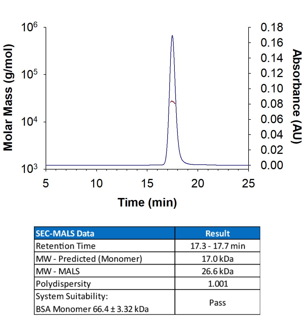
Recombinant Human PD‑1 His-tagged Protein SEC-MALS.
Recombinant Human PD-1 (Catalog # 8986-PD) has a molecular weight (MW) of 26.6 kDa as analyzed by SEC-MALS, suggesting that this protein is a monomer. MW may differ from predicted MW due to post-translational modifications (PTMs) present (i.e. Glycosylation).Binding of Human PD-1 to PD-L1/B7-H1 by surface plasmon resonance (SPR).
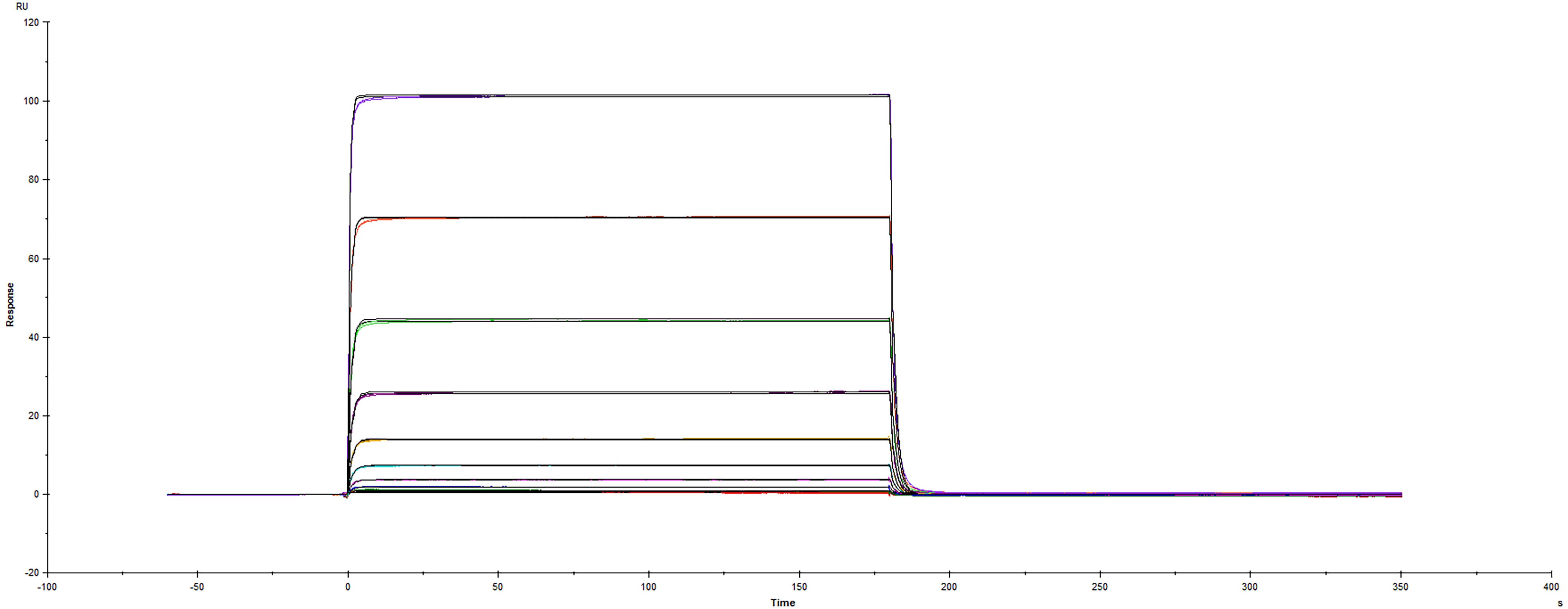
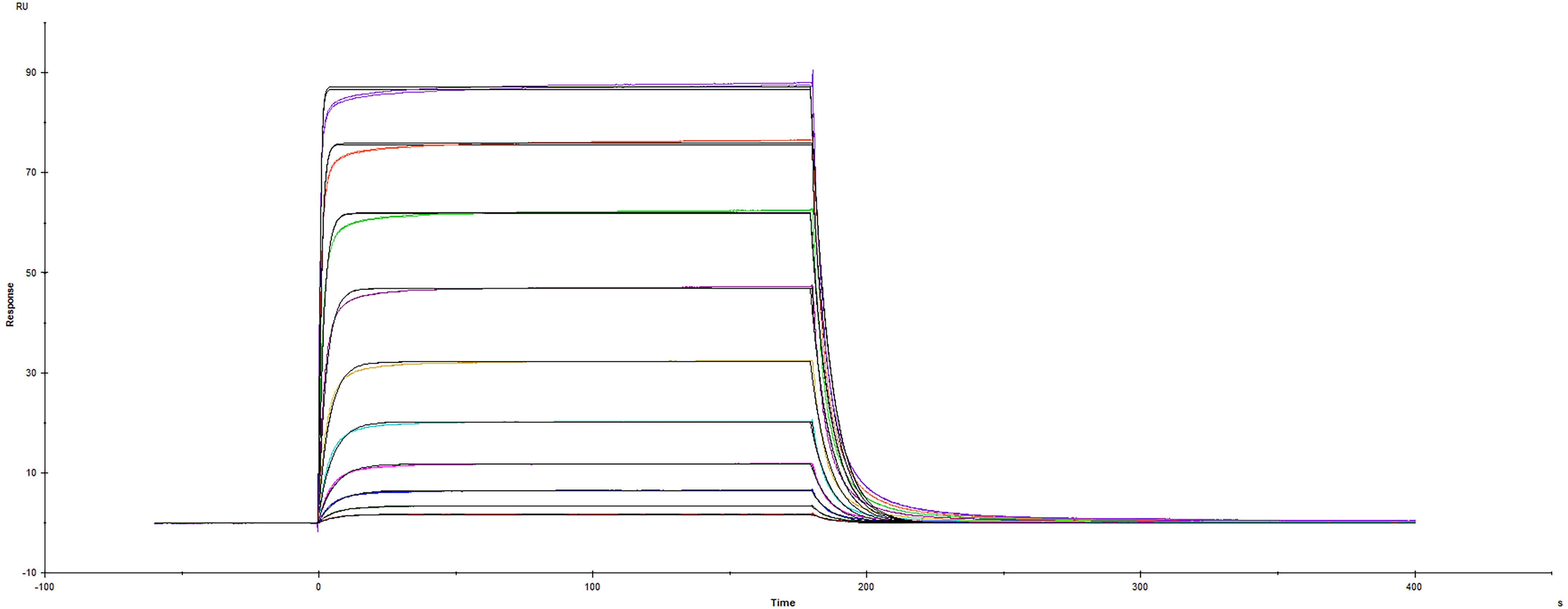
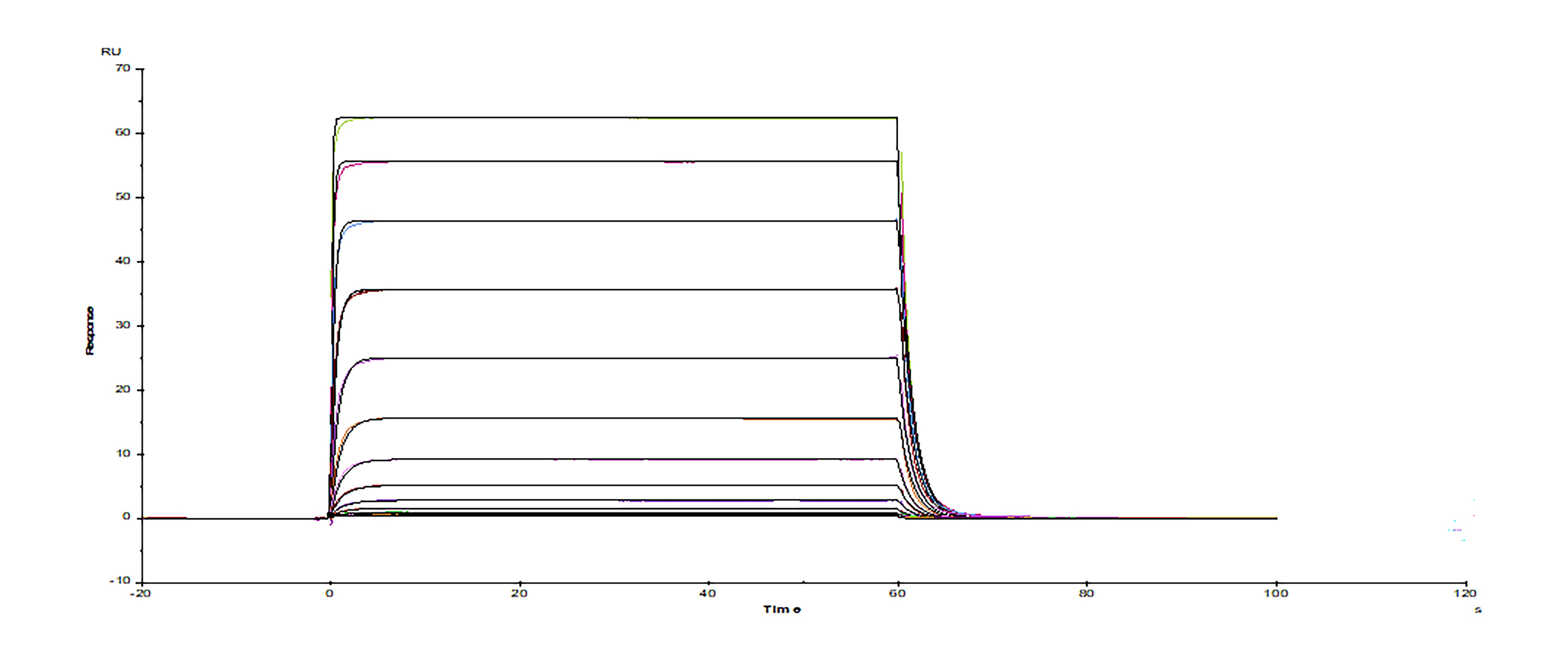
Recombinant Human PD-L1/B7-H1 Fc protein (156-B7) was immobilized on a Biacore Sensor Chip CM5, and binding to Recombinant Human PD-1 His protein (Catalog # 8986-PD) was measured at a concentration range between 6.0 nM and 3.05 uM. The double-referenced sensorgram was fit to a 1:1 binding model to determine the binding kinetics and affinity, with an affinity constant of KD=1.70 uM.Formulation, Preparation and Storage
8986-PD
| Formulation | Lyophilized from a 0.2 μm filtered solution in PBS. |
| Reconstitution |
Reconstitute at 400 μg/mL in PBS.
|
| Shipping | The product is shipped at ambient temperature. Upon receipt, store it immediately at the temperature recommended below. |
| Stability & Storage | Use a manual defrost freezer and avoid repeated freeze-thaw cycles.
|
Background: PD-1
References
- Ishida, Y. et al. (1992) EMBO J. 11:3887.
- Sharpe, A.H. and G. J. Freeman (2002) Nat. Rev. Immunol. 2:116.
- Coyle, A. and J. Gutierrez-Ramos (2001) Nat. Immunol. 2:203.
- Nishimura, H. and T. Honjo (2001) Trends Immunol. 22:265.
- Watanabe, N et al. (2003) Nat. Immunol. 4:670.
- Zhang, X. et al. (2004) Immunity 20:337.
- Lázár-Molnár, E. et al. (2008) Proc. Natl. Acad. Sci. USA 105:10483.
- Nishimura, H et al. (1996) Int. Immunol. 8:773.
- Keir, M.E. et al. (2008) Annu. Rev. Immunol. 26:677.
- Butte, M.J. et al. (2007) Immunity 27:111.
- Okazaki, T. et al. (2013) Nat. Immunol. 14:1212.
- Iwai, Y. et al. (2002) Proc. Natl. Acad. Sci. USA 99: 12293.
- Nogrady, B. (2014) Nature 513:S10.
- Swaika, A. et al. (2015) Mol. Immunol. 67: 4
Long Name
Programmed Death-1
Alternate Names
CD279, PD1, PDCD1, SLEB2
Entrez Gene IDs
Gene Symbol
PDCD1
UniProt
Additional PD-1 Products
Product Documents for Recombinant Human PD-1 His Tagged Protein, CF
Product Specific Notices for Recombinant Human PD-1 His Tagged Protein, CF
For research use only
Loading...
Loading...
Loading...


