
M1 Macrophage Flow Cytometry Panel
R&D Systems, part of Bio-Techne | Catalog # FMC-P-006
M1 Macrophage Flow Cytometry Panel

Key Product Details
Features
Save 10%, compared to buying individual components, by choosing our comprehensive Flow Panel!
Unlock Personalized Savings: Reach out to your sales representative to receive a discount on your custom panel!
Species
Human
Conjugate
Multiple
Scientific Data Images for M1 Macrophage Flow Cytometry Panel
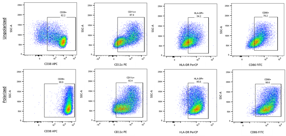
Multicolor flow cytometry panel to identify M1 Macrophages.
Monocytes were isolated from PBMCs via adherence depletion for 5 hours. Non-adherent cells were removed. Adherent cells were incubated for 6 days in C3 +10% huAB media with 50 ng/ml Recombinant Human GM-CSF (215-GM), media and cytokines are refreshed on day 3. For M1 polarization, 24 hours prior to staining, cells are incubated with 50 ng/ml human GM-CSF (215-GM), 50 ng/ml Recombinant Human IFN-gamma (285-IF), and 50 ng/ml LPS. Cells were stained with Anti-human CD3 Alexa Fluor ® 405, CD20 Alexa Fluor 405, CD56 Alexa Fluor 700, CD86 FITC, HLA-DR PerCP, CD11c PE,and CD38 APC. Cells were previously gated on Live CD3-CD20-CD56-. Find Staining Protocol Here.Formulation, Preparation, and Storage
Shipping
The product is shipped at ambient temperature. Upon receipt, store it immediately at the temperature recommended below.
Storage
Store the unopened product at 2 - 8 °C. Do not use past expiration date.
Background: Flow Cytometry Panel
Long Name
Flow Cytometry Panel
Additional Flow Cytometry Panel Products
Product Documents for M1 Macrophage Flow Cytometry Panel
Product Specific Notices for M1 Macrophage Flow Cytometry Panel
For research use only
Loading...
Loading...
Loading...