
TGF-beta 3 Antibody
R&D Systems, part of Bio-Techne | Catalog # MAB243


Scientific images may be resized, cropped, or adjusted for brightness or contrast for presentation purposes. These updates are limited to presentation and do not affect the underlying scientific interpretation of the data. Where available, additional source data may be provided upon request.
Conjugate
Catalog #
Key Product Details
Species Reactivity
Validated:
Multi-Species
Cited:
Human, Mouse, Avian - Chicken, Avian - Coturnix japonica (Quail), Canine
Applications
Validated:
Immunohistochemistry, Western Blot, Neutralization
Cited:
Immunohistochemistry, Western Blot, Neutralization, ELISA Development
Label
Unconjugated
Antibody Source
Monoclonal Mouse IgG1 Clone # 20724
Product Specifications
Immunogen
Spodoptera frugiperda, Sf 21 (baculovirus) derived recombinant human TGF-beta 3
Ala301-Ser412 (Tyr340Phe)
Accession # P10600
Ala301-Ser412 (Tyr340Phe)
Accession # P10600
Specificity
Detects TGF-beta 3 from multiple species in direct ELISAs and Western blots. In Western blots, less than 25% cross-reactivity with recombinant human (rh) TGF‑ beta1.2 and rhTGF-beta 2 is observed, and less than 2% cross‑reactivity with recombinant amphibian TGF-beta 5 and recombinant human TGF-beta 1 is observed. Neutralizes the biological activity of TGF-beta 3 but not TGF-beta 1, TGF-beta 2, or TGF-beta 5.
Clonality
Monoclonal
Host
Mouse
Isotype
IgG1
Endotoxin Level
<0.10 EU per 1 μg of the antibody by the LAL method.
Scientific Data Images for TGF-beta 3 Antibody
TGF-beta 3 Inhibition of IL-4-dependent Cell Proliferation and Neutralization by TGF-beta 3 Antibody.
TGF beta3 Antibody (Catalog # MAB243) neutralizes Recombinant Human TGF-beta 3 (243-B3) inhibition of Recombinant Mouse IL-4 (404-ML) induced proliferation in the HT-2 mouse T cell line. The Neutralization Dose (ND50) is typically 0.100-1.00 µg/mL.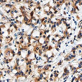
TGF‑ beta3 in Human Breast Cancer Tissue.
TGF-beta 3 was detected in immersion fixed paraffin-embedded sections of human breast cancer tissue using Mouse Anti-TGF-beta 3 Monoclonal Antibody (Catalog # MAB243) at 5 µg/mL overnight at 4 °C. Tissue was stained using the Anti-Mouse HRP-DAB Cell & Tissue Staining Kit (brown; CTS002) and counterstained with hematoxylin (blue). Specific staining was localized to cytoplasm in cancer cells. View our protocol for Chromogenic IHC Staining of Paraffin-embedded Tissue Sections.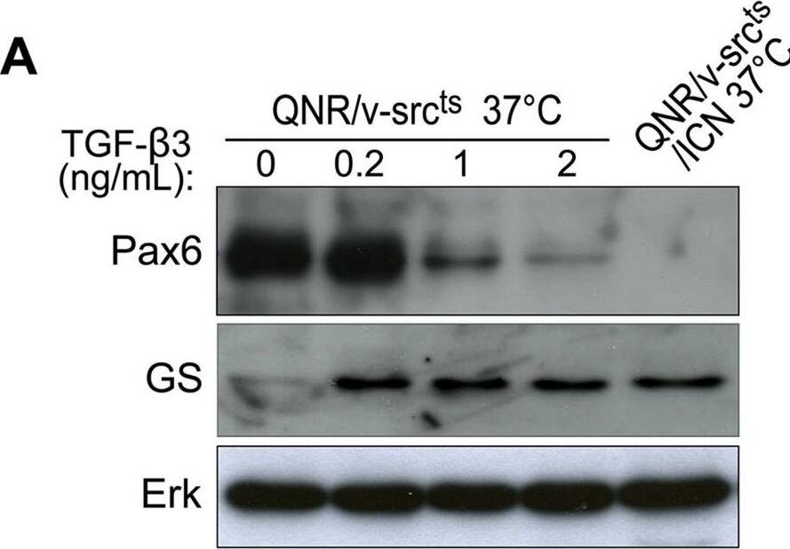
Detection of Coturnix japonica TGF-beta 3 by Western Blot
TGF-beta signaling controls commitment into glial differentiation.(A) QNR/v-srcts cells were treated at 37°C with increasing concentrations (0.2–2 ng/ml) of recombinant TGF-beta 3 protein during 7 days. Pax6 and Glutamine Synthetase (GS) were detected by western-blot. Protein loading was normalized using Erk antiserum. (B) Pax6 expression was analyzed by immunofluorescence after treatment of QNR/v-Srcts/ICN cells at 37°C with DMSO (control) or 10 µM SB431542 during 7 days. The majority of control QNR/v-Srcts/ICN cells did not express nuclear Pax6 but we observed a weak peri-nuclear labeling. Magnification x40. Image collected and cropped by CiteAb from the following open publication (https://pubmed.ncbi.nlm.nih.gov/21042581), licensed under a CC-BY license. Not internally tested by R&D Systems.Applications for TGF-beta 3 Antibody
Application
Recommended Usage
Immunohistochemistry
8-25 µg/mL
Sample: Immersion fixed paraffin-embedded sections of human breast cancer tissue
Sample: Immersion fixed paraffin-embedded sections of human breast cancer tissue
Western Blot
1 µg/mL
Sample: Recombinant Human TGF-beta 3 (Catalog # 243-B3) under non-reducing conditions only
Sample: Recombinant Human TGF-beta 3 (Catalog # 243-B3) under non-reducing conditions only
Neutralization
Measured by its ability to neutralize TGF‑ beta3 inhibition of IL‑4-dependent proliferation in the HT‑2 mouse T cell line. Tsang, M. et al. (1995) Cytokine 7:389. The Neutralization Dose (ND50) is typically 0.100-1.00 µg/mL in the presence of 0.1 ng/mL Recombinant Human TGF‑ beta3 and 7.5 ng/mL Recombinant Mouse IL‑4.
Formulation, Preparation, and Storage
Purification
Protein A or G purified from ascites
Reconstitution
Reconstitute at 0.5 mg/mL in sterile PBS. For liquid material, refer to CoA for concentration.
Formulation
Lyophilized from a 0.2 μm filtered solution in PBS with Trehalose. See Certificate of Analysis for details.
*Small pack size (-SP) is supplied either lyophilized or as a 0.2 µm filtered solution in PBS.
*Small pack size (-SP) is supplied either lyophilized or as a 0.2 µm filtered solution in PBS.
Shipping
Lyophilized product is shipped at ambient temperature. Liquid small pack size (-SP) is shipped with polar packs. Upon receipt, store immediately at the temperature recommended below.
Stability & Storage
Use a manual defrost freezer and avoid repeated freeze-thaw cycles.
- 12 months from date of receipt, -20 to -70 °C as supplied.
- 1 month, 2 to 8 °C under sterile conditions after reconstitution.
- 6 months, -20 to -70 °C under sterile conditions after reconstitution.
Background: TGF-beta 3
References
- Sporn, M.B. (2006) Cytokine Growth Factor Rev. 17:3.
- Dunker, N. and K. Krieglstein (2000) Eur. J. Biochem. 267:6982.
- Wahl, S.M. (2006) Immunol. Rev. 213:213.
- Chang, H. et al. (2002) Endocr. Rev. 23:787.
- Lin, J.S. et al. (2006) Reproduction 132:179.
- Hinck, A.P. et al. (1996) Biochemistry 35:8517.
- Mittl, P.R.E. et al. (1996) Protein Sci. 5:1261.
- Derynck, R. et al. (1988) EMBO J. 7:3737.
- Miyazono, K. et al. (1988) J. Biol. Chem. 263:6407.
- Oklu, R. and R. Hesketh (2000) Biochem. J. 352:601.
- de Caestecker, M. et al. (2004) Cytokine Growth Factor Rev. 15:1.
- Zuniga, J.E. et al. (2005) J. Mol. Biol. 354:1052.
Long Name
Transforming Growth Factor beta 3
Alternate Names
ARVD1, LDS5, RNHF, TGFB3, TGFbeta 3
Gene Symbol
TGFB3
UniProt
Additional TGF-beta 3 Products
Product Documents for TGF-beta 3 Antibody
Product Specific Notices for TGF-beta 3 Antibody
For research use only
Loading...
Loading...
Loading...
Loading...
Loading...