
Mouse/Rat Wnt-5a Antibody
R&D Systems, part of Bio-Techne | Catalog # AF645

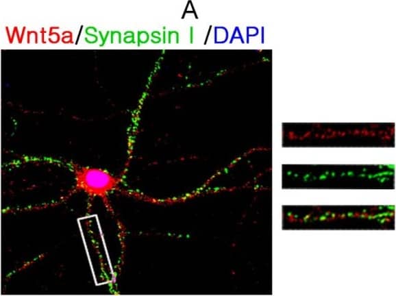

Scientific images may be resized, cropped, or adjusted for brightness or contrast for presentation purposes. These updates are limited to presentation and do not affect the underlying scientific interpretation of the data. Where available, additional source data may be provided upon request.
Conjugate
Catalog #
Key Product Details
Validated by
Biological Validation
Species Reactivity
Validated:
Mouse, Rat
Cited:
Human, Mouse, Rat, Frog - Xenopus (African Clawed Frog)
Applications
Validated:
Immunohistochemistry, Western Blot
Cited:
Immunohistochemistry, Immunohistochemistry-Paraffin, Immunohistochemistry-Frozen, Western Blot, Neutralization, Immunocytochemistry, Bioassay, Blocking, ELISA Detection, Microarray, Immunodepletion
Label
Unconjugated
Antibody Source
Polyclonal Goat IgG
Product Specifications
Immunogen
E. coli-derived recombinant mouse Wnt-5a peptide
Gln254-Cys334
Accession # P22725
Gln254-Cys334
Accession # P22725
Specificity
Detects mouse and rat Wnt-5a in direct ELISAs and Western blots. In direct ELISAs, approximately 5% cross-reactivity with recombinant mouse (rm) Wnt‑5b is observed and less than 2% cross-reactivity with rmWnt-1, rmWnt-3a, rmWnt-4, rmWnt-11, and rmWnt-16 is observed.
Clonality
Polyclonal
Host
Goat
Isotype
IgG
Scientific Data Images for Mouse/Rat Wnt-5a Antibody
Wnt-5a in Mouse Embryonic Rib.
Wnt-5a was detected in immersion fixed paraffin-embedded sections of mouse embryonic rib using 15 µg/mL Mouse/Rat Wnt-5a Antigen Affinity-purified Polyclonal Antibody (Catalog # AF645) overnight at 4 °C. Tissue was stained with the Anti-Goat HRP-DAB Cell & Tissue Staining Kit (brown; CTS008) and counterstained with hematoxylin (blue). View our protocol for Chromogenic IHC Staining of Paraffin-embedded Tissue Sections.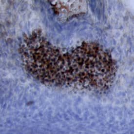
Wnt‑5a in Mouse Embryo.
Wnt-5a was detected in immersion fixed frozen sections of mouse embryo using Mouse/Rat Wnt-5a Antigen Affinity-purified Polyclonal Antibody (Catalog # AF645) at 15 µg/mL overnight at 4 °C. Tissue was stained using the Anti-Goat HRP-DAB Cell & Tissue Staining Kit (brown; CTS008) and counterstained with hematoxylin (blue). View our protocol for Chromogenic IHC Staining of Frozen Tissue Sections.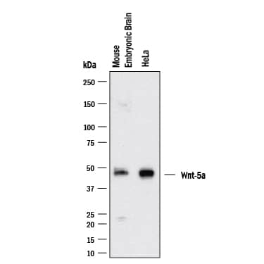
Detection of Mouse/Rat Wnt-5a by Western Blot.
Western blot shows lysates of HeLa Human Cervical Epithelial Carcinoma Cells and Mouse Brain (embryo E14). PVDF membrane was probed with 2 µg/mL of Goat Anti-Mouse/Rat Wnt-5a Antigen Affinity-purified Polyclonal Antibody (Catalog # AF645) followed by HRP-conjugated Anti-Goat IgG Secondary Antibody (Catalog # HAF017). A specific band was detected for Wnt-5a at approximately ~42kDa kDa (as indicated). This experiment was conducted under reducing conditions and using Western Blot Buffer Group 1.


Applications for Mouse/Rat Wnt-5a Antibody
Application
Recommended Usage
Immunohistochemistry
5-15 µg/mL
Sample: Immersion fixed paraffin-embedded sections of mouse embryonic rib and immersion fixed frozen sections of mouse embryo
Sample: Immersion fixed paraffin-embedded sections of mouse embryonic rib and immersion fixed frozen sections of mouse embryo
Western Blot
2 µg/mL
Sample: Lysates of HeLa Human Cervical Epithelial Carcinoma Cells and Mouse Brain (embryo E14)
Sample: Lysates of HeLa Human Cervical Epithelial Carcinoma Cells and Mouse Brain (embryo E14)
Reviewed Applications
Read 6 reviews rated 3.5 using AF645 in the following applications:
Formulation, Preparation, and Storage
Purification
Antigen Affinity-purified
Reconstitution
Reconstitute at 0.2 mg/mL in sterile PBS. For liquid material, refer to CoA for concentration.
Formulation
Lyophilized from a 0.2 μm filtered solution in PBS with Trehalose. *Small pack size (SP) is supplied either lyophilized or as a 0.2 µm filtered solution in PBS.
Shipping
Lyophilized product is shipped at ambient temperature. Liquid small pack size (-SP) is shipped with polar packs. Upon receipt, store immediately at the temperature recommended below.
Stability & Storage
Use a manual defrost freezer and avoid repeated freeze-thaw cycles.
- 12 months from date of receipt, -20 to -70 °C as supplied.
- 1 month, 2 to 8 °C under sterile conditions after reconstitution.
- 6 months, -20 to -70 °C under sterile conditions after reconstitution.
Background: Wnt-5a
Wnt proteins are secreted glycoproteins that contain a conserved pattern of 23-24 cysteine residues. Wnts play critical roles in both carcinogenesis and embryonic development for a variety of organisms. Wnts bind to receptors of the Frizzled family, sometimes in conjunction with other membrane-associated proteins such as LRPs or proteoglycans. Downstream effects of Wnt signaling occur through different intracellular components, depending on which pathway is activated. Three pathways have been characterized: the canonical Wnt/ beta-catenin pathway, the Wnt/Ca2+ pathway, and the planar cell polarity (1, 2).
Wnt-5a is part of the subgroup of Wnts that are not axis-inducing in Xenopus embryos and do not transform C57MG mammary epithelial cells. This subgroup is also implicated in the Wnt/Ca2+ pathway, playing roles in cell movements and cell adhesion (3). This non-canonical Wnt pathway can inhibit canonical Wnt/ beta-catenin signaling. In Wnt-5a deficient mouse embryos, beta-catenin accumulates in the limb bud suggesting that Wnt-5a normally promotes degradation of beta-catenin (4). Likewise, in Xenopus embryos Wnt-5a antagonizes the ability of the canonical Wnt subgroup to induce a secondary axis (5). Wnt-5a is implicated in various types of cancer and has complex roles. It acts as a tumor suppressor for mammary, B-cell, colon, and uroepithelial cancer cells but is up-regulated in melanomas, where expression levels correlate with severity of metastasis (3). Furthermore, aberrant Wnt-5a signaling results in other diseases such as rheumatoid arthritis (6). Like other developmental growth factors Wnt-5a has diverse roles in development. They are too numerous to enunciate here, as functions span from early anterior-posterior development and gastrulation movements to maintaining hematopoietic stem cell population, lung morphogenesis, and limb outgrowth. Mouse and human Wnt-5a share 97% amino acid identity.
References
- Miller, J.R. (2002) Genome Biol. 3:3001.
- Roelink, H. and R. Nusse (1991) Genes Dev. 5:381.
- Veeman, M.T. et al. (2003) Developmental Cell 5:367.
- Topol, L. et al. (2003) J. Cell Biol 162:899.
- Torres, M. et al. (1996) J. Cell Biol. 133:1123.
- Sen, M. et al. (2001) Arthritis & Rheumatism 44:772.
Long Name
Wingless-type MMTV Integration Site Family, Member 5a
Alternate Names
Wnt5a
Gene Symbol
WNT5A
UniProt
Additional Wnt-5a Products
Product Documents for Mouse/Rat Wnt-5a Antibody
Product Specific Notices for Mouse/Rat Wnt-5a Antibody
For research use only
Loading...
Loading...
Loading...
Loading...
Loading...