Scientific Data Examples for Mouse Podoplanin Antibody
Multiplex Immunofluorescence
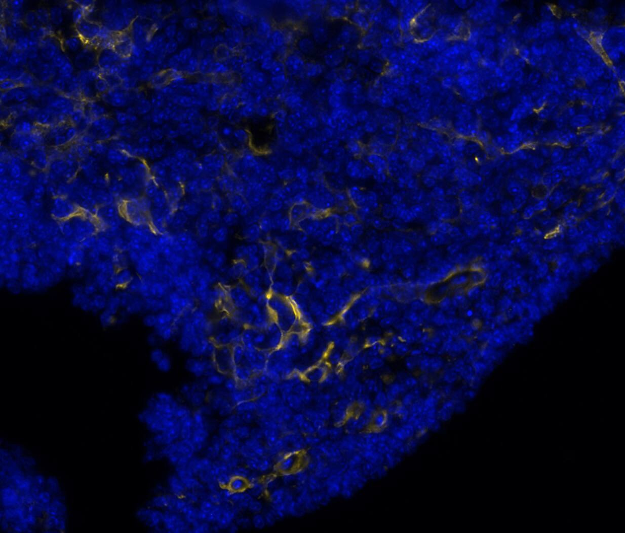
Detection of PDPN in Mouse Thymus via seqIF™ staining on COMET™
PDPN was detected in immersion fixed paraffin-embedded sections of mouse thymus using Rabbit Anti-Mouse PDPN Monoclonal Antibody (Catalog # MAB11813) at 0.1ug/mL at 37 ° Celsius for 4 minutes. Before incubation with the primary antibody, tissue underwent an all-in-one dewaxing and antigen retrieval preprocessing using PreTreatment Module (PT Module) and Dewax and HIER Buffer H (pH 9; Epredia Catalog # TA-999-DHBH). Tissue was stained using the Alexa Fluor™ Plus 555 Goat anti-Rabbit IgG Secondary Antibody at 1:100 at 37 ° Celsius for 2 minutes. (Yellow; Lunaphore Catalog # DR555RB) and counterstained with DAPI (blue; Lunaphore Catalog # DR100). Specific staining was localized to the membrane. Protocol available in COMET™ Panel Builder.
Multiplex Immunofluorescence
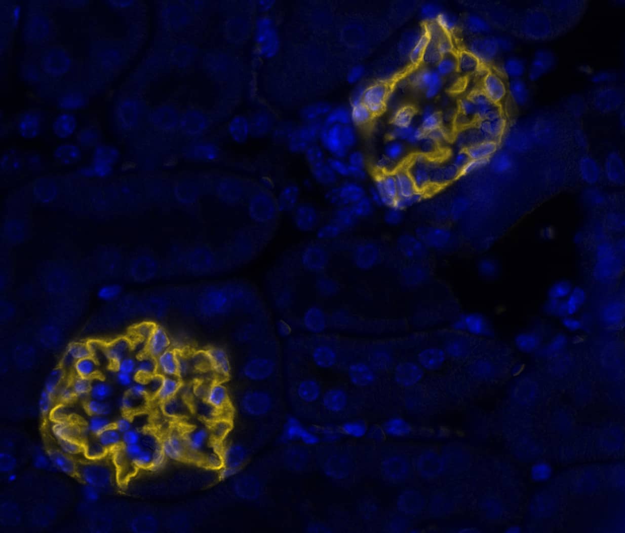
Detection of PDPN in Mouse Kidney via seqIF™ staining on COMET™
PDPN was detected in immersion fixed paraffin-embedded sections of mouse kidney using Rabbit Anti-Mouse PDPN Monoclonal Antibody (Catalog # MAB11813) at 0.1 μg/mL at 37 ° Celsius for 4 minutes. Before incubation with the primary antibody, tissue underwent an all-in-one dewaxing and antigen retrieval preprocessing using PreTreatment Module (PT Module) and Dewax and HIER Buffer H (pH 9; Epredia Catalog # TA-999-DHBH). Tissue was stained using the Alexa Fluor™ Plus 555 Goat anti-Rabbit IgG Secondary Antibody at 1:100 at 37 ° Celsius for 2 minutes. (Yellow; Lunaphore Catalog # DR555RB) and counterstained with DAPI (blue; Lunaphore Catalog # DR100). Specific staining was localized to the membrane. Protocol available in COMET™ Panel Builder.
Multiplex Immunofluorescence
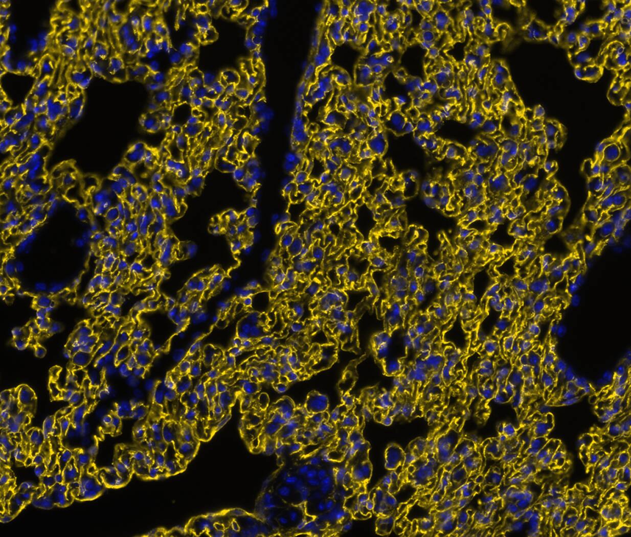
Detection of PDPN in Mouse Lung via seqIF™ staining on COMET™
PDPN was detected in immersion fixed paraffin-embedded sections of mouse lung using Rabbit Anti-Mouse PDPN Monoclonal Antibody (Catalog # MAB11813) at 0.1ug/mL at 37 ° Celsius for 4 minutes. Before incubation with the primary antibody, tissue underwent an all-in-one dewaxing and antigen retrieval preprocessing using PreTreatment Module (PT Module) and Dewax and HIER Buffer H (pH 9; Epredia Catalog # TA-999-DHBH). Tissue was stained using the Alexa Fluor™ Plus 555 Goat anti-Rabbit IgG Secondary Antibody at 1:100 at 37 ° Celsius for 2 minutes. (Yellow; Lunaphore Catalog # DR555RB) and counterstained with DAPI (blue; Lunaphore Catalog # DR100). Specific staining was localized to the membrane. Protocol available in COMET™ Panel Builder.
Multiplex Immunofluorescence
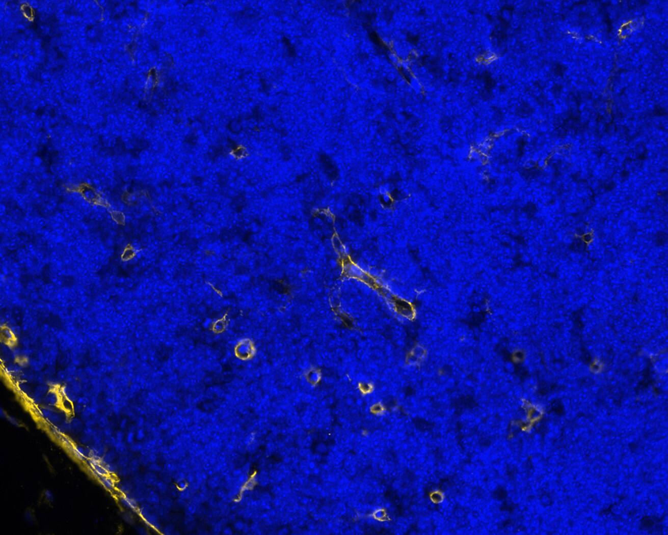
Detection of PDPN in Mouse Thymus via seqIF™ staining on COMET™
PDPN was detected in immersion fixed paraffin-embedded sections of mouse thymus using Rabbit Anti-Mouse PDPN Monoclonal Antibody (Catalog # MAB11813) at 0.5ug/mL at 37 ° Celsius for 4 minutes. Before incubation with the primary antibody, tissue underwent an all-in-one dewaxing and antigen retrieval preprocessing using PreTreatment Module (PT Module) and Dewax and HIER Buffer H (pH 9; Epredia Catalog # TA-999-DHBH). Tissue was stained using the Alexa Fluor™ Plus 647 Goat anti-Rabbit IgG Secondary Antibody at 1:200 at 37 ° Celsius for 2 minutes. (Yellow; Lunaphore Catalog # DR647RB) and counterstained with DAPI (blue; Lunaphore Catalog # DR100). Specific staining was localized to the membrane. Protocol available in COMET™ Panel Builder.
Western Blot
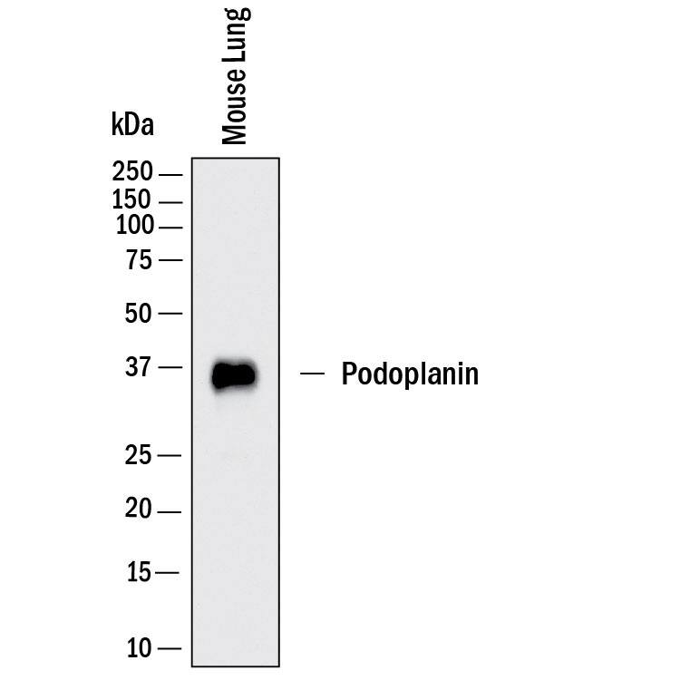
Detection of Mouse Podoplanin by Western Blot.
Western Blot shows lysates of mouse lung. PVDF membrane was probed with 0.1 µg/ml of Rabbit Anti-Mouse Podoplanin Monoclonal Antibody (Catalog # MAB11813) followed by HRP-conjugated Anti-Rabbit IgG Secondary Antibody (Catalog # HAF008). A specific band was detected for Podoplanin at approximately 36 kDa (as indicated). This experiment was conducted under reducing conditions and using Western Blot Buffer Group 1.
Immunohistochemistry
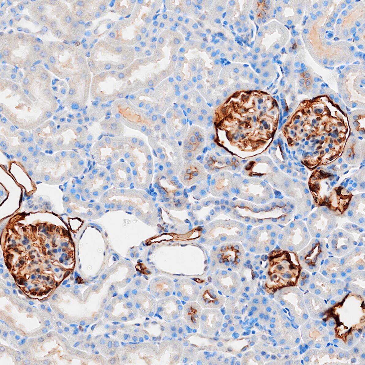
Immersion fixed paraffin-embedded sections of mouse kidney
Podoplanin was detected in immersion fixed paraffin-embedded sections of mouse kidney using Rabbit Anti-Mouse Podoplanin Monoclonal Antibody (Catalog # MAB11813) at 1 µg/ml overnight at 4 °C. Before incubation with the primary antibody, tissue was subjected to heat-induced epitope retrieval using VisUCyte Antigen Retrieval Reagent-Basic (Catalog # VCTS021). Tissue was stained using the HRP-conjugated Anti-Rabbit IgG Secondary Antibody (Catalog # HAF008) and counterstained with hematoxylin (blue). Specific staining was localized to the membrane. View our protocol for Chromogenic IHC Staining of Paraffin-embedded Tissue Sections.
Formulation, Preparation and Storage
Purification
Protein A or G purified from hybridoma culture supernatant
Reconstitution
Reconstitute lyophilized material at 0.2 mg/ml in sterile PBS. For liquid material, refer to CoA for concentration.
Formulation
Lyophilized from a 0.2 μm filtered solution in PBS with Trehalose.
Shipping
Lyophilized product is shipped at ambient temperature. Liquid small pack size (-SP) is shipped with polar packs. Upon receipt, store immediately at the temperature recommended below.
Stability & Storage
Use a manual defrost freezer and avoid repeated freeze-thaw cycles.
12 months from date of receipt, -20 to -70 °C as supplied.
1 month, 2 to 8 °C under sterile conditions after reconstitution.
6 months, -20 to -70 °C under sterile conditions after reconstitution.
Reconstitution Calculator
Background: Podoplanin
Podoplanin, also known as T1 alpha (T1 alpha ), is a mucin type transmembrane glycoprotein with extensive O-glycosylation. It is specifically expressed by lymphatic endothelial cells but not blood vascular endothelial cells. In addition, non-endothelial cells in numerous normal tissues also express the protein. Within the region used as the immunogen, mouse Podoplanin shares 73.5% and 29% amino acid sequence homology with rat and human Podoplanin, respectively.
Item
Value
Long Name
Lung Type-I Cell Membrane-associated Glycoprotein, Isoform A