
Mouse IL-23R PE-conjugated Antibody
R&D Systems, part of Bio-Techne | Catalog # FAB16861P


Conjugate
Catalog #
Key Product Details
Validated by
Biological Validation
Species Reactivity
Validated:
Mouse
Cited:
Mouse
Applications
Validated:
Flow Cytometry
Cited:
Flow Cytometry
Label
Phycoerythrin (Excitation = 488 nm, Emission = 565-605 nm)
Antibody Source
Monoclonal Rat IgG1 Clone # 753317
Product Specifications
Immunogen
Mouse myeloma cell line NS0-derived recombinant mouse IL-23 R
Gly24-Asp372
Accession # Q8K4B4
Gly24-Asp372
Accession # Q8K4B4
Specificity
Detects endogenous mouse IL-23 R by flow cytometry.
Clonality
Monoclonal
Host
Rat
Isotype
IgG1
Scientific Data Images for Mouse IL-23R PE-conjugated Antibody
Detection of IL‑23 R in Mouse Splenocytes by Flow Cytometry.
Mouse splenocytes treated with 10 µg/mL Anti-CD3 (Catalog # MAB484), 5 µg/mL Anti-CD28 (Catalog # AF483), 10 ng/mL Recombinant Mouse TGF-beta 1 (Catalog # 100-B), 20 ng/mL Recombinant Mouse IL-23 (Catalog # 1887-ML), 40 ng/mL Recombinant Mouse IL-6 (Catalog # 406-ML), and 10 ng/mL Recombinant Mouse IL-1 beta (Catalog # 401-ML) for 5 days to induce T17 activation were stained with Rat Anti-Mouse CD4 APC-conjugated Monoclonal Antibody (Catalog # FAB554A) and either (A) Rat Anti-Mouse IL-23 R PE-conjugated Monoclonal Antibody (Catalog # FAB16861P) or (B) Rat IgG1Phycoerythrin Isotype Control (Catalog # IC005P). View our protocol for Staining Membrane-associated Proteins.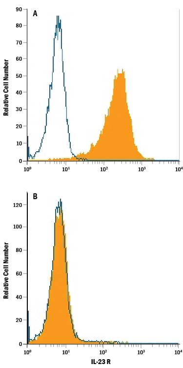
Detection of IL-23 R in HEK293 Human Cell Line Transfected with Mouse IL-23 R by Flow Cytometry.
HEK293 human embryonic kidney cell line transfected with (A) mouse IL-23 R or (B) irrelevant transfectants was stained with Rat Anti-Mouse IL-23 R PE-conjugated Monoclonal Antibody (Catalog # FAB16861P, filled histogram) or isotype control antibody (Catalog # IC005P, open histogram). View our protocol for Staining Membrane-associated Proteins.Applications for Mouse IL-23R PE-conjugated Antibody
Application
Recommended Usage
Flow Cytometry
0.25-1 µg/106 cells
Sample: Mouse splenocytes treated with Anti-CD3 (Catalog # MAB484), Anti-CD28 (Catalog # AF483), Recombinant Mouse TGF-beta 1 (Catalog # 100-B), Recombinant Mouse IL-23 (Catalog # 1887-ML), Recombinant Mouse IL-6 (Catalog # 406-ML), and Recombinant Mouse IL-1 beta (Catalog # 401-ML) to induce T17 activation or HEK293 human embryonic kidney cell line transfected with mouse IL-23 R
Sample: Mouse splenocytes treated with Anti-CD3 (Catalog # MAB484), Anti-CD28 (Catalog # AF483), Recombinant Mouse TGF-beta 1 (Catalog # 100-B), Recombinant Mouse IL-23 (Catalog # 1887-ML), Recombinant Mouse IL-6 (Catalog # 406-ML), and Recombinant Mouse IL-1 beta (Catalog # 401-ML) to induce T17 activation or HEK293 human embryonic kidney cell line transfected with mouse IL-23 R
Spectra Viewer
Plan Your Experiments
Use our spectra viewer to interactively plan your experiments, assessing multiplexing options. View the excitation and emission spectra for our fluorescent dye range and other commonly used dyes.
Spectra Viewer
Formulation, Preparation, and Storage
Purification
Protein A or G purified from hybridoma culture supernatant
Formulation
Supplied in a saline solution containing BSA and Sodium Azide.
Shipping
The product is shipped with polar packs. Upon receipt, store it immediately at the temperature recommended below.
Stability & Storage
Protect from light. Do not freeze.
- 12 months from date of receipt, 2 to 8 °C as supplied.
Background: IL-23R
References
- Oppmann, B. et al. (2000) Immunity 13:715.
- Lankford, C.S. and D.M. Frucht (2003) J. Leukoc. Biol. 73:49.
- Parham, C. et al. (2002) J. Immunol. 168:5448.
- Belladonna, M.L. et al. (2002) J. Immunol. 168:5448.
- Aggarwal, S. et al. (2003) J. Biol. Chem. 278:1910.
Long Name
Interleukin 23 Receptor
Alternate Names
IL-23 R, IL23R
Gene Symbol
IL23R
UniProt
Additional IL-23R Products
Product Documents for Mouse IL-23R PE-conjugated Antibody
Product Specific Notices for Mouse IL-23R PE-conjugated Antibody
For research use only
Loading...
Loading...
Loading...
Loading...
Loading...