
Human Tie-1 PE-conjugated Antibody
R&D Systems, part of Bio-Techne | Catalog # FAB619P

Scientific images may be resized, cropped, or adjusted for brightness or contrast for presentation purposes. These updates are limited to presentation and do not affect the underlying scientific interpretation of the data. Where available, additional source data may be provided upon request.
Conjugate
Catalog #
Key Product Details
Species Reactivity
Validated:
Human
Cited:
Human
Applications
Validated:
Flow Cytometry
Cited:
Flow Cytometry, Immunocytochemistry
Label
Phycoerythrin (Excitation = 488 nm, Emission = 565-605 nm)
Antibody Source
Polyclonal Goat IgG
Product Specifications
Immunogen
Mouse myeloma cell line NS0-derived recombinant human Tie-1
Ala22-Gln760
Accession # P35590
Ala22-Gln760
Accession # P35590
Specificity
Detects human Tie-1 in direct ELISAs and Western blots. In direct ELISAs, less than 1% cross-reactivity with recombinant human Tie‑2 is observed.
Clonality
Polyclonal
Host
Goat
Isotype
IgG
Scientific Data Images for Human Tie-1 PE-conjugated Antibody
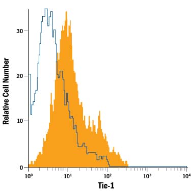
Detection of Tie‑1 in Human Umbilical Vein Endothelial Cells (HUVEC) by Flow Cytometry.
Human umbilical vein endothelial cells were stained with Goat Anti-Human Tie-1 PE-conjugated Antigen Affinity-purified Polyclonal Antibody (Catalog # FAB619P, filled histogram) or isotype control antibody (Catalog # IC108P, open histogram). View our protocol for Staining Membrane-associated Proteins.Applications for Human Tie-1 PE-conjugated Antibody
Application
Recommended Usage
Flow Cytometry
10 µL/106 cells
Sample: Human umbilical vein endothelial cells (HUVEC)
Sample: Human umbilical vein endothelial cells (HUVEC)
Spectra Viewer
Plan Your Experiments
Use our spectra viewer to interactively plan your experiments, assessing multiplexing options. View the excitation and emission spectra for our fluorescent dye range and other commonly used dyes.
Spectra Viewer
Formulation, Preparation, and Storage
Purification
Antigen Affinity-purified
Formulation
Supplied in a saline solution containing BSA and Sodium Azide.
Shipping
The product is shipped with polar packs. Upon receipt, store it immediately at the temperature recommended below.
Stability & Storage
Protect from light. Do not freeze.
- 12 months from date of receipt, 2 to 8 °C as supplied.
Background: Tie-1
Tie-1/Tie (tyrosine kinase with Ig and EGF homology domains 1) and Tie-2/Tek comprise a receptor tyrosine kinase (RTK) subfamily with unique structural characteristics: two immunoglobulin-like domains, flanking three epidermal growth factor (EGF)-like domains followed by three fibronectin type III-like repeats in the extracellular region and a split tyrosine kinase domain in the cytoplasmic region. These receptors are expressed primarily on endothelial and hematopoietic progenitor cells and play critical roles in angiogenesis, vasculogenesis and hematopoiesis.
Human Tie-1 cDNA encodes a 1138 amino acid (aa) residue precursor protein with a 24 residue putative signal peptide, a 735 residue extracellular domain, a 25 residue transmembrane domain, and a 354 residue cytoplasmic domain. Ligands which bind and activate Tie-1 have not been identified. Based on gene-targeting studies, the in vivo functions of Tie-1 have been shown to be related to endothelial cell differentiation and the maintenance of integrity of the endothelium.
References
- Partanen, J. and D.J. Dumont (1999) Curr. Top. Microbiol. Immunol. 237:159
- Sato, T.N. et al. (1995) Nature 376:70
Long Name
Tyrosine Kinase with Immunoglobulin and Epidermal Growth Factor Homology Domains 1
Alternate Names
Tie1
Gene Symbol
TIE1
UniProt
Additional Tie-1 Products
Product Documents for Human Tie-1 PE-conjugated Antibody
Product Specific Notices for Human Tie-1 PE-conjugated Antibody
For research use only
Loading...
Loading...
Loading...
Loading...
Loading...