
Human SOX21 Antibody
R&D Systems, part of Bio-Techne | Catalog # AF3538



Scientific images may be resized, cropped, or adjusted for brightness or contrast for presentation purposes. These updates are limited to presentation and do not affect the underlying scientific interpretation of the data. Where available, additional source data may be provided upon request.
Conjugate
Catalog #
Key Product Details
Species Reactivity
Validated:
Human
Cited:
Human, Mouse
Applications
Validated:
Immunohistochemistry, Western Blot, Immunocytochemistry
Cited:
Immunohistochemistry, Immunofluorescence, Immunoprecipitation
Label
Unconjugated
Antibody Source
Polyclonal Goat IgG
Product Specifications
Immunogen
E. coli-derived recombinant human SOX21
Ser2-Leu276 (Gly102Ser)
Accession # Q9Y651
Ser2-Leu276 (Gly102Ser)
Accession # Q9Y651
Specificity
Detects human SOX21 in direct ELISAs and Western blots. In direct ELISAs and Western blots, less than 1% cross-reactivity with recombinant human (rh) SOX1, rhSOX2 and rhSOX15 is observed.
Clonality
Polyclonal
Host
Goat
Isotype
IgG
Scientific Data Images for Human SOX21 Antibody
SOX21 in NTera‑2 Human Cell Line.
SOX21 was detected in immersion fixed NTera-2 human testicular embryonic carcinoma cell line using Goat Anti-Human SOX21 Antigen Affinity-purified Polyclonal Antibody (Catalog # AF3538) at 10 µg/mL for 3 hours at room temperature. Cells were stained using the NorthernLights™ 557-conjugated Anti-Goat IgG Secondary Antibody (red; NL001) and counterstained with DAPI (blue). Specific staining was localized to nuclei. View our protocol for Fluorescent ICC Staining of Cells on Coverslips.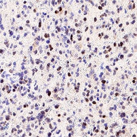
Detection of SOX21 in Human Glioblastoma.
SOX21 was detected in immersion fixed paraffin-embedded sections of human glioblastoma using Goat Anti-Human SOX21 Antigen Affinity-purified Polyclonal Antibody (Catalog # AF3538) at 0.5 µg/ml for 1 hour at room temperature followed by incubation with the HRP-conjugated Anti-Goat IgG Secondary Antibody (Catalog # HAF017) or the Anti-Goat IgG VisUCyte™ HRP Polymer Antibody (Catalog # VC004). Before incubation with the primary antibody, tissue was subjected to heat-induced epitope retrieval using VisUCyte Antigen Retrieval Reagent-Basic (Catalog # VCTS021). Tissue was stained using DAB (brown) and counterstained with hematoxylin (blue). Specific staining was localized to the nucleus. View our protocol for Chromogenic IHC Staining of Paraffin-embedded Tissue Sections.
Detection of SOX21 by Flow Cytometry
Transcriptomic impact of SOX21 expression in GPCs.Volcano plots displaying differential gene expression induced by SOX21 in JM11 (A) and JM13 (B) GPCs after 48 hours. Genes significantly upregulated (red) and downregulated (blue) are shown (false discovery rate < 0.01). C Heatmaps show gene sets and associated Gene Ontology (GO) terms deregulated upon SOX21 induction. D, E Enrichment analysis of SOX motifs (black line) and AP-1 motifs (green line) within SOX21 ChIP-seq peaks detected in JM11 (D) and JM13 (E) GPCs, with motif distances to the center of SOX21 peaks measured in base pairs (bp). P-values of best-matching SOX and AP-1 motifs are shown. F Enrichment of transcription factor binding motifs across SOX21 ChIP-seq regions in comparison with non-targeted control chromatin regions. Graphs illustrating the most significant spacing distances between centrally enriched SOX and AP-1 motifs in SOX21 ChIP-seq peaks detected in JM11 (G) and JM13 (H) GPCs. The gap between motifs is based on the spacing between the last nucleotide in the SOX motif and the first nucleotide in AP-1. I, J Co-immunoprecipitation assays showing interactions between FLAG-tagged SOX21 and c-JUN in JM11 and JM13 GPCs treated with DNase I. K Genomic mapping of c-JUN ChIP-seq reads within SOX21 ChIP-seq peak regions detected in JM11 and JM13 GPCs. L, M Genomic regions surrounding CAMK2D (L) and ADRA1D (M) genes, illustrating ChIP-seq binding profiles for c-JUN, SOX21, and SOX2 in JM11 GPCs. Image collected and cropped by CiteAb from the following open publication (https://www.nature.com/articles/s41419-026-08442-5), licensed under a CC-BY license. Not internally tested by R&D Systems.


Applications for Human SOX21 Antibody
Application
Recommended Usage
Immunocytochemistry
5-15 µg/mL
Sample: Immersion fixed NTera‑2 human testicular embryonic carcinoma cell line
Sample: Immersion fixed NTera‑2 human testicular embryonic carcinoma cell line
Immunohistochemistry
5-15 µg/mL
Sample: Immersion fixed paraffin-embedded sections of human glioblastoma
Sample: Immersion fixed paraffin-embedded sections of human glioblastoma
Western Blot
0.1 µg/mL
Sample: Recombinant Human SOX21
Sample: Recombinant Human SOX21
Formulation, Preparation, and Storage
Purification
Antigen Affinity-purified
Reconstitution
Reconstitute at 0.2 mg/mL in sterile PBS. For liquid material, refer to CoA for concentration.
Formulation
Lyophilized from a 0.2 μm filtered solution in PBS with Trehalose. See Certificate of Analysis for details.
*Small pack size (-SP) is supplied either lyophilized or as a 0.2 µm filtered solution in PBS.
*Small pack size (-SP) is supplied either lyophilized or as a 0.2 µm filtered solution in PBS.
Shipping
Lyophilized product is shipped at ambient temperature. Liquid small pack size (-SP) is shipped with polar packs. Upon receipt, store immediately at the temperature recommended below.
Stability & Storage
Use a manual defrost freezer and avoid repeated freeze-thaw cycles.
- 12 months from date of receipt, -20 to -70 °C as supplied.
- 1 month, 2 to 8 °C under sterile conditions after reconstitution.
- 6 months, -20 to -70 °C under sterile conditions after reconstitution.
Background: SOX21
Long Name
Transcription Factor SOX21
Alternate Names
SOX25
Gene Symbol
SOX21
UniProt
Additional SOX21 Products
Product Documents for Human SOX21 Antibody
Product Specific Notices for Human SOX21 Antibody
For research use only
Loading...
Loading...
Loading...
Loading...
Loading...