
Human/Mouse/Rat FBPase 1 Antibody
R&D Systems, part of Bio-Techne | Catalog # AF7285

Conjugate
Catalog #
Key Product Details
Species Reactivity
Human, Mouse, Rat
Applications
Immunohistochemistry, Western Blot
Label
Unconjugated
Antibody Source
Polyclonal Sheep IgG
Product Specifications
Immunogen
E. coli-derived recombinant human FBPase 1
Ala2-Gln338 (Arg218Lys)
Accession # P09467
Ala2-Gln338 (Arg218Lys)
Accession # P09467
Specificity
Detects human, mouse, rat FBPase 1 in Western blots and detects human FBPase 1 in direct ELISAs.
Clonality
Polyclonal
Host
Sheep
Isotype
IgG
Scientific Data Images for Human/Mouse/Rat FBPase 1 Antibody
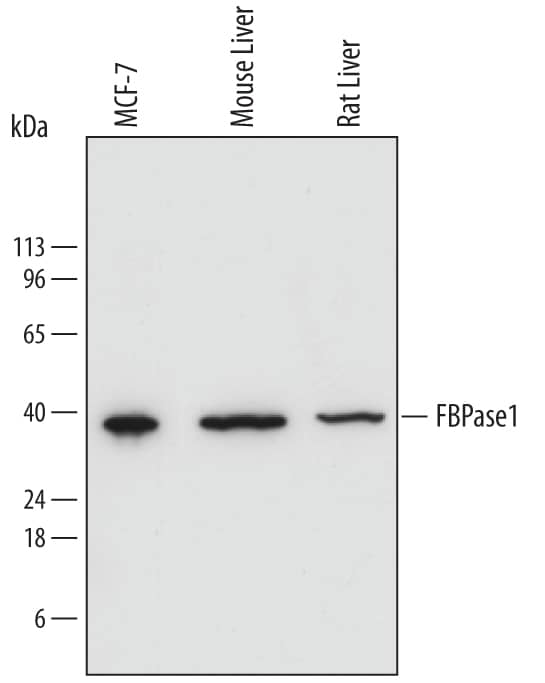
Detection of Human, Mouse, and Rat FBPase 1 by Western Blot.
Western blot shows lysates of MCF-7 human breast cancer cell line, mouse liver tissue, and rat liver tissue. PVDF membrane was probed with 0.2 µg/mL of Sheep Anti-Human/Mouse/Rat FBPase 1 Antigen Affinity-purified Polyclonal Antibody (Catalog # AF7285) followed by HRP-conjugated Anti-Sheep IgG Secondary Antibody (Catalog # HAF016). A specific band was detected for FBPase 1 at approximately 37 kDa (as indicated). This experiment was conducted under reducing conditions and using Immunoblot Buffer Group 1.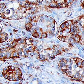
FBPase 1 in Human Liver Cancer Tissue.
FBPase 1 was detected in immersion fixed paraffin-embedded sections of human liver cancer tissue using Sheep Anti-Human/Mouse/Rat FBPase 1 Antigen Affinity-purified Polyclonal Antibody (Catalog # AF7285) at 15 µg/mL for 1 hour at room temperature followed by incubation with the Anti-Sheep IgG VisUCyte™ HRP Polymer Antibody (Catalog # VC006). Tissue was stained using DAB (brown) and counterstained with hematoxylin (blue). Specific staining was localized to cytoplasm in cancer cells. View our protocol for IHC Staining with VisUCyte HRP Polymer Detection Reagents.Applications for Human/Mouse/Rat FBPase 1 Antibody
Application
Recommended Usage
Immunohistochemistry
5-15 µg/mL
Sample: Immersion fixed paraffin-embedded sections of human liver cancer tissue
Sample: Immersion fixed paraffin-embedded sections of human liver cancer tissue
Western Blot
0.2 µg/mL
Sample: MCF‑7 human breast cancer cell line, mouse liver tissue, and rat liver tissue
Sample: MCF‑7 human breast cancer cell line, mouse liver tissue, and rat liver tissue
Formulation, Preparation, and Storage
Purification
Antigen Affinity-purified
Reconstitution
Sterile PBS to a final concentration of 0.2 mg/mL. For liquid material, refer to CoA for concentration.
Formulation
Lyophilized from a 0.2 μm filtered solution in PBS with Trehalose. *Small pack size (SP) is supplied either lyophilized or as a 0.2 µm filtered solution in PBS.
Shipping
Lyophilized product is shipped at ambient temperature. Liquid small pack size (-SP) is shipped with polar packs. Upon receipt, store immediately at the temperature recommended below.
Stability & Storage
Use a manual defrost freezer and avoid repeated freeze-thaw cycles.
- 12 months from date of receipt, -20 to -70 °C as supplied.
- 1 month, 2 to 8 °C under sterile conditions after reconstitution.
- 6 months, -20 to -70 °C under sterile conditions after reconstitution.
Background: FBPase 1
FBPase 1 (Fructose 1,6 bisphosphatase 1 [Fru-1,6-P2ase]; also FBP-1 phosphohydrolase 1) is a 35-37 kDa member of the FBPase Class I family of enzymes. It is widely expressed, being found in cells diverse as monocytes, hepatocytes, pancreatic beta-cells and striated (skeletal plus cardiac) muscle cells. FBPase 1 converts Fru-1,6-P2 into Fru-6-P, a molecule that is subsequently used to generate glucose. Human FBPase 1 is 338 amino acids (aa) in length. It contains one catalytic region (aa 18-332) and an NLS between aa 203-208. FBPase 1 exhibits divalent cation dependency, and exists as a homotetramer within the cell. Full-length human FBPase 1 shares 85% aa sequence identity with mouse FBPase 1.
Long Name
Fructose-1,6-bisphosphatase 1
Alternate Names
FBP1
Gene Symbol
FBP1
UniProt
Additional FBPase 1 Products
Product Documents for Human/Mouse/Rat FBPase 1 Antibody
Product Specific Notices for Human/Mouse/Rat FBPase 1 Antibody
For research use only
Loading...
Loading...
Loading...
Loading...
Loading...