
Human/Mouse Meprin beta Subunit/MEP1B Antibody
R&D Systems, part of Bio-Techne | Catalog # MAB28951

Scientific images may be resized, cropped, or adjusted for brightness or contrast for presentation purposes. These updates are limited to presentation and do not affect the underlying scientific interpretation of the data. Where available, additional source data may be provided upon request.
Conjugate
Catalog #
Key Product Details
Species Reactivity
Validated:
Human, Mouse
Cited:
Porcine
Applications
Validated:
Immunohistochemistry, Western Blot
Cited:
Immunohistochemistry, Western Blot
Label
Unconjugated
Antibody Source
Monoclonal Rat IgG1 Clone # 289731
Product Specifications
Immunogen
Human embryonic kidney cell line HEK293-derived recombinant human Meprin beta Subunit/MEP1B
Thr23-Ser293
Accession # Q16820
Thr23-Ser293
Accession # Q16820
Specificity
Detects the pro form of human or mouse Meprin beta Subunit/MEP1B in Western blots. It does not react with the mature form.
Clonality
Monoclonal
Host
Rat
Isotype
IgG1
Scientific Data Images for Human/Mouse Meprin beta Subunit/MEP1B Antibody
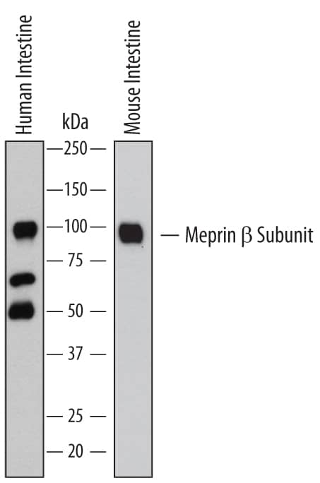
Detection of Human and Mouse Meprin beta Subunit/ MEP1B by Western Blot.
Western blot shows lysates of human intestine tissue and mouse intestine tissue. PVDF membrane was probed with 1 µg/mL of Rat Anti-Human/Mouse Meprin beta Subunit/MEP1B Monoclonal Antibody (Catalog # MAB28951) followed by HRP-conjugated Anti-Rat IgG Secondary Antibody (Catalog # HAF005). A specific band was detected for Meprin beta Subunit/MEP1B at approximately 97 kDa (as indicated). This experiment was conducted under reducing conditions and using Immunoblot Buffer Group 1.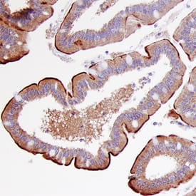
Meprin beta Subunit/MEP1B in Human Intestine.
Meprin beta Subunit/MEP1B was detected in immersion fixed paraffin-embedded sections of human intestine using Human/Mouse Meprin beta Subunit/MEP1B Monoclonal Antibody (Catalog # MAB28951) at 1 µg/mL overnight at 4 °C. Before incubation with the primary antibody, tissue was subjected to heat-induced epitope retrieval using Antigen Retrieval Reagent-Basic (Catalog # CTS013). Tissue was stained using the Anti-Rat HRP-DAB Cell & Tissue Staining Kit (brown; Catalog # CTS017) and counterstained with hemotoxylin (blue). Specific staining was localized to the Brush border of epithelial cells. View our protocol for Chromogenic IHC Staining of Paraffin-embedded Tissue Sections. This application has not been tested in mouse samples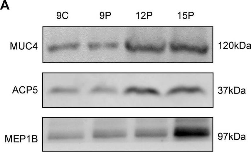
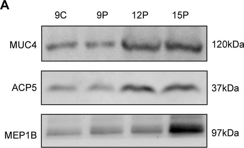
Detection of Meprin beta Subunit/MEP1B by Western Blot
Representative proteins in ULF-EVs whose abundance increased with pregnancy development also significantly increased after ULF-EVs treatment of pTr2 cells. A Western blot analysis of MUC4, ACP5 and MEP1B in ULF-EVs. B Relative abundance of MUC4, ACP5 and MEP1B in pTr2 cells treated with ULF-EVs. C Images of the embryo-maternal interface stained with MEP1B antibodies. MEP1B was obviously expressed in the endometrium and trophoblast. The scale bar indicates 100 nm. D Quantitative analysis of MEP1B by assessing the average integrated optical density (IOD) in the endometrium. The data are displayed as mean ± SD and different lowercase letters correspond to significant differences at the p < 0.05 threshold. E Quantitative analysis of MEP1B by assessing IOD in the trophoblast. Asterisks indicate significant differences (mean ± SD) between 12 and 15P (**p < 0.01). F The relative expression level of MEP1B mRNA was determined by qRT-PCR. The data are displayed as mean ± SD and asterisk indicate significant differences (*p < 0.05) Image collected and cropped by CiteAb from the following open publication (https://pubmed.ncbi.nlm.nih.gov/36882792), licensed under a CC-BY license. Not internally tested by R&D Systems.Applications for Human/Mouse Meprin beta Subunit/MEP1B Antibody
Application
Recommended Usage
Immunohistochemistry
8-25 µg/mL
Sample: Immersion fixed paraffin-embedded sections of human intestine
Sample: Immersion fixed paraffin-embedded sections of human intestine
Western Blot
1 µg/mL
Sample: Human intestine tissue and mouse intestine tissue
Sample: Human intestine tissue and mouse intestine tissue
Formulation, Preparation, and Storage
Purification
Protein A or G purified from hybridoma culture supernatant
Reconstitution
Reconstitute at 0.5 mg/mL in sterile PBS. For liquid material, refer to CoA for concentration.
Formulation
Lyophilized from a 0.2 μm filtered solution in PBS with Trehalose. *Small pack size (SP) is supplied either lyophilized or as a 0.2 µm filtered solution in PBS.
Shipping
Lyophilized product is shipped at ambient temperature. Liquid small pack size (-SP) is shipped with polar packs. Upon receipt, store immediately at the temperature recommended below.
Stability & Storage
Use a manual defrost freezer and avoid repeated freeze-thaw cycles.
- 12 months from date of receipt, -20 to -70 °C as supplied.
- 1 month, 2 to 8 °C under sterile conditions after reconstitution.
- 6 months, -20 to -70 °C under sterile conditions after reconstitution.
Background: Meprin beta Subunit/MEP1B
Meprins are multimeric proteases composed of alpha and beta subunits, which are members of the astacin family of zinc endopeptidases (1, 2). Both subunits form disulfide-linked homo- or heterooligomers, which are also referred to as Meprin A (composed of alpha subunits with or without beta subunits) and Merpin B (composed of beta subunits only) (3). Although the two subunits share 42% amino acid (aa) sequence identity, they differ significantly in their oligomeric structure, post-translational processing and subsequently cellular location, and substrate and peptide bond specificity (4). The 701 aa sequence of human Merpin beta subunit precursor consists of a signal peptide (aa 1 to 21), a pro region (aa 22 to 61), and a mature chain (aa 62 to 701) containing catalytic (aa 62 to 259), MAM (aa 260 to 429), MATH (aa 430 to 585), EGF-like (aa 604 to 644), transmembrane (aa 653 to 673), and cytoplasmic (aa 674 to 701) domains.
References
- Bond, J.S. and R.J. Beynon (1995) Protein Sci. 4:1247.
- Stocker, W. et al. (1995) Protein Sci. 4:823.
- Bertenshaw, G.P. et al. (2001) J. Biol. Chem. 276:13248.
- Ishmael, F.T. et al. (2005) J. Biol. Chem. 280:13895.
Alternate Names
MEP1B
Gene Symbol
MEP1B
UniProt
Additional Meprin beta Subunit/MEP1B Products
Product Documents for Human/Mouse Meprin beta Subunit/MEP1B Antibody
Product Specific Notices for Human/Mouse Meprin beta Subunit/MEP1B Antibody
For research use only
Loading...
Loading...
Loading...
Loading...
Loading...