
Human ErbB2/Her2 Antibody
R&D Systems, part of Bio-Techne | Catalog # AF1129


Scientific images may be resized, cropped, or adjusted for brightness or contrast for presentation purposes. These updates are limited to presentation and do not affect the underlying scientific interpretation of the data. Where available, additional source data may be provided upon request.
Conjugate
Catalog #
Key Product Details
Species Reactivity
Validated:
Human
Cited:
Human, Transgenic Mouse
Applications
Validated:
Immunohistochemistry, Western Blot, Inhibition of Cell Growth, Flow Cytometry, CyTOF-ready
Cited:
Western Blot, Flow Cytometry, Immunocytochemistry, Immunoprecipitation, ELISA Capture, ELISA Development
Label
Unconjugated
Antibody Source
Polyclonal Goat IgG
Product Specifications
Immunogen
Mouse myeloma cell line NS0-derived recombinant human ErbB2/Her2
Thr23-Thr652
Accession # P04626
Thr23-Thr652
Accession # P04626
Specificity
Detects human ErbB2/Her2 in direct ELISAs and Western blots. In direct ELISAs, less than 20% cross-reactivity with recombinant mouse ErbB2 is observed and less than 5% cross-reactivity with recombinant human (rh) ErbB3, rhErbB4, and rhEGF R is observed.
Clonality
Polyclonal
Host
Goat
Isotype
IgG
Endotoxin Level
<0.10 EU per 1 μg of the antibody by the LAL method.
Scientific Data Images for Human ErbB2/Her2 Antibody
ErbB2/Her2 in Human Breast Cancer Tissue.
ErbB2/Her2 was detected in immersion fixed paraffin-embedded sections of human breast cancer tissue using Goat Anti-Human ErbB2/Her2 Antigen Affinity-purified Polyclonal Antibody (Catalog # AF1129) at 1.7 µg/mL overnight at 4 °C. Tissue was stained using the Anti-Goat HRP-DAB Cell & Tissue Staining Kit (brown; Catalog # CTS008) and counterstained with hematoxylin (blue). Specific staining was localized to epithelial cells. View our protocol for Chromogenic IHC Staining of Paraffin-embedded Tissue Sections.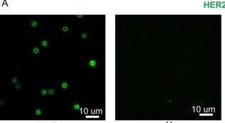
Detection of ErbB2/Her2 by Immunocytochemistry/ Immunofluorescence
Characterization of tEVs isolated breast cancer (BC) patient serum using iSUF. (A) Left. Characteristic fluorescence images of HER2, CD24, miR21, CD63, and CD9 expression on isolated EVs immobilized on magnetic beads from BC patients and healthy donors. Right. Fluorescence intensity histograms of HER2, CD24, miR21, CD63, and CD9 of isolated EVs for BC patients (n = 10) and healthy donors (n = 14). (B) Total fluorescence intensity quantification of HER2, CD24, miR21, CD63, and CD9 expression level on isolated EVs from BC patients and healthy donors. Patients demonstrated higher expression levels of HER2, CD24, and miR21 than healthy donors (p < 0.05). Patients and HDs demonstrated similar expression level of CD63 and CD9 (p > 0.05). P patient; H healthy donor. Image collected and cropped by CiteAb from the following open publication (https://pubmed.ncbi.nlm.nih.gov/33850163), licensed under a CC-BY license. Not internally tested by R&D Systems.Applications for Human ErbB2/Her2 Antibody
Application
Recommended Usage
CyTOF-ready
Ready to be labeled using established conjugation methods. No BSA or other carrier proteins that could interfere with conjugation.
Flow Cytometry
2.5 µg/106 cells
Sample: MCF-7 human breast cancer cell line
Sample: MCF-7 human breast cancer cell line
Immunohistochemistry
5-15 µg/mL
Sample: Immersion fixed paraffin-embedded sections of human breast cancer tissue
Sample: Immersion fixed paraffin-embedded sections of human breast cancer tissue
Inhibition of Cell Growth
Measured by its ability to inhibit proliferation in the SK-BR-3 human breast cancer cell line. Brodowicz, T. et al. (1997) Int. J. Cancer 73:875. The ED50 for this effect is typically 0.06-0.3 µg/mL.
Western Blot
0.1 µg/mL
Sample: Recombinant Human ErbB2/Her2 Fc Chimera (Catalog # 1129-ER)
Sample: Recombinant Human ErbB2/Her2 Fc Chimera (Catalog # 1129-ER)
Reviewed Applications
Read 3 reviews rated 4 using AF1129 in the following applications:
Formulation, Preparation, and Storage
Purification
Antigen Affinity-purified
Reconstitution
Reconstitute at 0.2 mg/mL in sterile PBS. For liquid material, refer to CoA for concentration.
Formulation
Lyophilized from a 0.2 μm filtered solution in PBS with Trehalose. *Small pack size (SP) is supplied either lyophilized or as a 0.2 µm filtered solution in PBS.
Shipping
Lyophilized product is shipped at ambient temperature. Liquid small pack size (-SP) is shipped with polar packs. Upon receipt, store immediately at the temperature recommended below.
Stability & Storage
Use a manual defrost freezer and avoid repeated freeze-thaw cycles.
- 12 months from date of receipt, -20 to -70 °C as supplied.
- 1 month, 2 to 8 °C under sterile conditions after reconstitution.
- 6 months, -20 to -70 °C under sterile conditions after reconstitution.
Background: ErbB2/Her2
ErbB2, also called Neu and Her2 (human epidermal growth factor receptor 2), is a type I membrane glycoprotein that is a member of the ErbB family of tyrosine kinase receptors. ErbB family members serve as receptors for the epidermal growth factor (EGF) family of growth factors. ErbB2 is widely expressed in epithelial cells and has also been found to be over-expressed in a large number of breast carcinomas. Among ErbB family members, ErbB2 is unique in that it has no identified ligands. Rather, ErbB2 heterodimerizes with the other members of the ErbB family (ErbB1 (EGFR), ErbB3, ErbB4) to form higher affinity signaling complexes. Because ErbB3 contains a defective kinase domain, the kinase domain of ErbB2 is responsible for initiating the tyrosine phosphorylation signal through the heterodimeric receptor. It has been found that a discrete three amino acid signal in the ErbB3 cytoplasmic domain is critical for transactivation of ErbB2. Interestingly, this same three amino acid signal has also been found in ErbB1 and ErbB4. Phosphoinositide 3-kinase has been shown to play a role in ErbB2 signal transduction. The cytoplasmic domain of ErbB2 has been shown to associate with beta-catenin and plakoglobin. Human ErbB2 consists of 1255 amino acids (aa) with a 21 aa signal sequence, a 631 aa extracellular domain, a 23 aa transmembrane region, and a 580 aa cytoplasmic domain. ErbB2 can be shed from the cell surface by proteolytic cleavage by an unidentified protease. ErbB2 appears to play roles in development, cancer, communication at the neuromuscular junction, and regulation of cell growth and differentiation (1-10).
References
- Coussens, L. et. al. (1985) Science 230:1132.
- Yamamoto, T. et. al. (1986) Nature 319:230.
- Kanai, Y. et. al. (1995) Biochem. Biophys. Res. Commun. 208:1067.
- Codony-Servat, J. et. al. (1999) Cancer Res. 59:1196.
- Carraway, K.L. 3rd et. al. (1994) J. Biol. Chem. 269:14303.
- Emkey, R. and C.R. Kahn (1997) J. Biol. Chem. 272:31172.
- Schaefer, G. et. al. (1999) J. Biol. Chem. 274:859.
- Schlessinger, J. (2000) Cell 103:211.
- Hellyer, N.J. et. al. (2001) J. Biol. Chem. 276:42153.
- Daly, R.J. (1999) Growth Factors 16:255.
Long Name
Receptor Tyrosine Protein Kinase ErbB2
Alternate Names
CD340, HER2, Neu Oncogene, NGL, TKR1
Gene Symbol
ERBB2
UniProt
Additional ErbB2/Her2 Products
Product Documents for Human ErbB2/Her2 Antibody
Product Specific Notices for Human ErbB2/Her2 Antibody
For research use only
Loading...
Loading...
Loading...
Loading...
Loading...
Loading...