
Human eIF2 alpha Antibody
R&D Systems, part of Bio-Techne | Catalog # AF3997

Scientific images may be resized, cropped, or adjusted for brightness or contrast for presentation purposes. These updates are limited to presentation and do not affect the underlying scientific interpretation of the data. Where available, additional source data may be provided upon request.
Conjugate
Catalog #
Key Product Details
Species Reactivity
Validated:
Human
Cited:
Human, Mouse
Applications
Validated:
Western Blot, Simple Western
Cited:
Western Blot
Label
Unconjugated
Antibody Source
Polyclonal Goat IgG
Product Specifications
Immunogen
E. coli-derived recombinant human eIF2 alpha
Met1-Asp315
Accession # Q53XC0
Met1-Asp315
Accession # Q53XC0
Specificity
Detects human, mouse, and rat eIF2 alpha in Western blots.
Clonality
Polyclonal
Host
Goat
Isotype
IgG
Scientific Data Images for Human eIF2 alpha Antibody
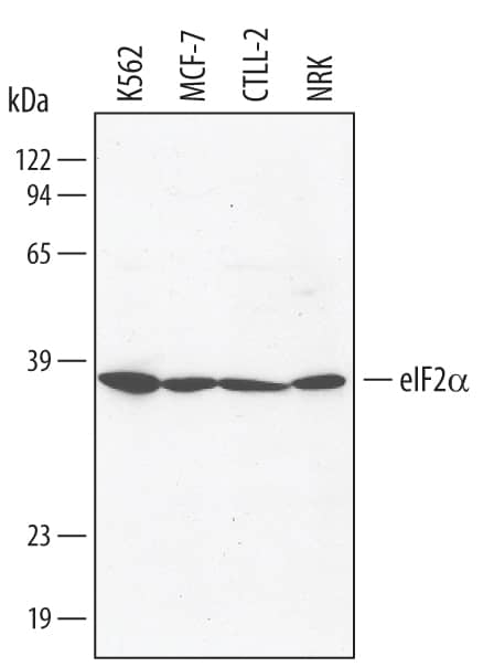
Detection of Human eIF2 alpha by Western Blot.
Western blot shows lysates of K562 human chronic myelogenous leukemia cell line, MCF-7 human breast cancer cell line, CTLL-2 mouse cytotoxic T cell line, and NRK rat normal kidney cell line. PVDF membrane was probed with 1 µg/mL of Goat Anti-Human eIF2a Antigen Affinity-purified Polyclonal Antibody (Catalog # AF3997) followed by HRP-conjugated Anti-Goat IgG Secondary Antibody (Catalog # HAF109). A specific band was detected for eIF2a at approximately 38 kDa (as indicated). This experiment was conducted under reducing conditions and using Immunoblot Buffer Group 1.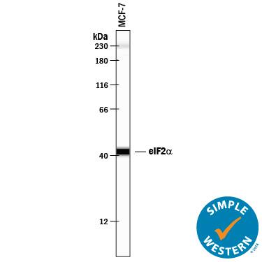
Detection of Human eIF2 alpha by Simple WesternTM.
Simple Western lane view shows lysates of MCF-7 human breast cancer cell line, loaded at 0.2 mg/mL. A specific band was detected for eIF2a at approximately 42 kDa (as indicated) using 10 µg/mL of Goat Anti-Human eIF2a Antigen Affinity-purified Polyclonal Antibody (Catalog # AF3997) followed by 1:50 dilution of HRP-conjugated Anti-Goat IgG Secondary Antibody (Catalog # HAF109). This experiment was conducted under reducing conditions and using the 12-230 kDa separation system. Non-specific interaction with the 230 kDa Simple Western standard may be seen with this antibody.
Detection of eIF2 alpha by Western Blot
Analysis of signal transduction during CHIKV infection in the presence of 50 nM silvestrol. (A) HEK 293T cells were seeded in six-well plates and were infected with CHIKV using an MOI of 1, and 50 nM silvestrol was added where indicated. Cells were treated with IFN alpha for 30 min before harvest if indicated, and Western blot analysis of cell lysates was performed. (−) Untreated cells; (+) treated cells. In lanes 3–6, cells were infected with CHIKV. The CHIKV E2 protein, STAT1, eIF2 alpha, and their phosphorylated proteins were detected with specific antibodies and secondary HRP-coupled antibodies, and the ECL detection system (Amersham, Freiburg). Equal loading of each blot was controlled by detection of beta-actin; and, (B) Uninfected HEK293T cells were treated with IFN alpha for 30 min before harvest and either treated with silvestrol for 16 h or left untreated. Western blot analysis of cell lysates was performed and p-STAT1 and STAT1 were detected. STAT1 served as a loading control. Image collected and cropped by CiteAb from the following open publication (https://pubmed.ncbi.nlm.nih.gov/30380742), licensed under a CC-BY license. Not internally tested by R&D Systems.Applications for Human eIF2 alpha Antibody
Application
Recommended Usage
Simple Western
10 µg/mL
Sample: MCF‑7 human breast cancer cell line
Sample: MCF‑7 human breast cancer cell line
Western Blot
1 µg/mL
Sample: K562 human chronic myelogenous leukemia cell line chronic myelogenous leukemia, MCF-7 human breast cancer cell liner, CTLL-2 mouse cytotoxic T cell line, and NRK rat normal kidney cells
Sample: K562 human chronic myelogenous leukemia cell line chronic myelogenous leukemia, MCF-7 human breast cancer cell liner, CTLL-2 mouse cytotoxic T cell line, and NRK rat normal kidney cells
Reviewed Applications
Read 2 reviews rated 5 using AF3997 in the following applications:
Formulation, Preparation, and Storage
Purification
Antigen Affinity-purified
Reconstitution
Reconstitute at 0.2 mg/mL in sterile PBS. For liquid material, refer to CoA for concentration.
Formulation
Lyophilized from a 0.2 μm filtered solution in PBS with Trehalose. *Small pack size (SP) is supplied either lyophilized or as a 0.2 µm filtered solution in PBS.
Shipping
Lyophilized product is shipped at ambient temperature. Liquid small pack size (-SP) is shipped with polar packs. Upon receipt, store immediately at the temperature recommended below.
Stability & Storage
Use a manual defrost freezer and avoid repeated freeze-thaw cycles.
- 12 months from date of receipt, -20 to -70 °C as supplied.
- 1 month, 2 to 8 °C under sterile conditions after reconstitution.
- 6 months, -20 to -70 °C under sterile conditions after reconstitution.
Background: eIF2 alpha
Long Name
Eukaryotic Translation Initiation Factor 2 Alpha
Alternate Names
EIF2S1
Gene Symbol
EIF2S1
UniProt
Additional eIF2 alpha Products
Product Documents for Human eIF2 alpha Antibody
Product Specific Notices for Human eIF2 alpha Antibody
For research use only
Loading...
Loading...
Loading...
Loading...
Loading...