
Human ECE-1 Antibody
R&D Systems, part of Bio-Techne | Catalog # AF1784

Scientific images may be resized, cropped, or adjusted for brightness or contrast for presentation purposes. These updates are limited to presentation and do not affect the underlying scientific interpretation of the data. Where available, additional source data may be provided upon request.
Conjugate
Catalog #
Key Product Details
Species Reactivity
Validated:
Human
Cited:
Human, Mouse, Rat
Applications
Validated:
Immunohistochemistry, Western Blot, Intracellular Staining by Flow Cytometry, Immunoprecipitation, CyTOF-ready
Cited:
Immunohistochemistry, Western Blot, Immunocytochemistry
Label
Unconjugated
Antibody Source
Polyclonal Goat IgG
Product Specifications
Immunogen
Mouse myeloma cell line NS0-derived recombinant human ECE-1
Gln90-Trp770
Accession # P42892
Gln90-Trp770
Accession # P42892
Specificity
Detects human ECE-1 in direct ELISAs and Western blots. In direct ELISAs and Western blots, approximately 5% cross-reactivity with recombinant human ECE-2 is observed.
Clonality
Polyclonal
Host
Goat
Isotype
IgG
Scientific Data Images for Human ECE-1 Antibody
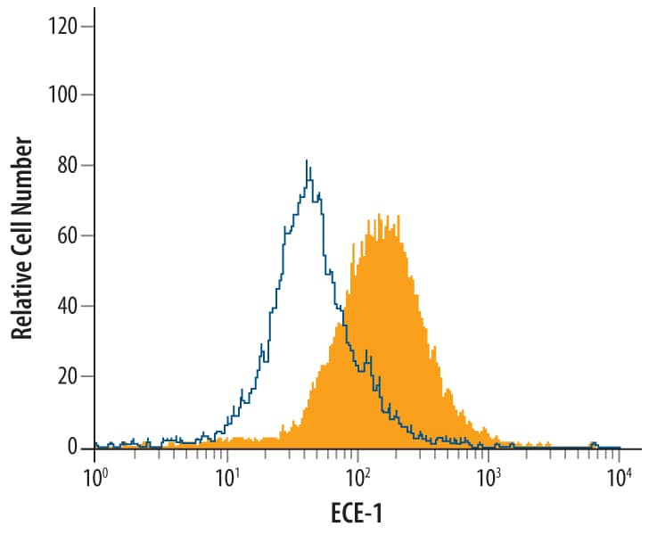
Detection of ECE-1 in MCF-7 Human Cell Line by Flow Cytometry.
MCF-7 human breast cancer cell line was stained with Human ECE-1 Antigen Affinity-purified Polyclonal Antibody (Catalog # AF1784, filled histogram) or control antibody (Catalog # AB-108-C, open histogram), followed by Allophycocyanin-conjugated Anti-Goat IgG Secondary Antibody (Catalog # F0108). To facilitate intracellular staining, cells were fixed with paraformaldehyde and permeabilized with saponin.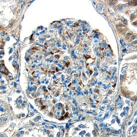
ECE-1 in Human Kidney.
ECE-1 was detected in immersion fixed paraffin-embedded sections of human kidney using Goat Anti-Human ECE-1 Antigen Affinity-purified Polyclonal Antibody (Catalog # AF1784) at 15 µg/mL overnight at 4 °C. Tissue was stained using the Anti-Goat HRP-DAB Cell & Tissue Staining Kit (brown; Catalog # CTS008) and counterstained with hematoxylin (blue). Specific labeling was localized to the endothelial cells in glomeruli. View our protocol for Chromogenic IHC Staining of Paraffin-embedded Tissue Sections.Applications for Human ECE-1 Antibody
Application
Recommended Usage
CyTOF-ready
Ready to be labeled using established conjugation methods. No BSA or other carrier proteins that could interfere with conjugation.
Immunohistochemistry
5-15 µg/mL
Sample: Immersion fixed paraffin-embedded sections of human kidney
Sample: Immersion fixed paraffin-embedded sections of human kidney
Immunoprecipitation
25 µg/mL
Sample: Conditioned cell culture medium spiked with Recombinant Human ECE‑1 (Catalog # 1784‑ZN), see our available Western blot detection antibodies
Sample: Conditioned cell culture medium spiked with Recombinant Human ECE‑1 (Catalog # 1784‑ZN), see our available Western blot detection antibodies
Intracellular Staining by Flow Cytometry
2.5 µg/106 cells
Sample: MCF-7 human breast cancer cell line fixed with paraformaldehyde and permeabilized with saponin
Sample: MCF-7 human breast cancer cell line fixed with paraformaldehyde and permeabilized with saponin
Western Blot
0.1 µg/mL
Sample: Recombinant Human ECE-1 (Catalog # 1784-ZN)
Sample: Recombinant Human ECE-1 (Catalog # 1784-ZN)
Formulation, Preparation, and Storage
Purification
Antigen Affinity-purified
Reconstitution
Reconstitute at 0.2 mg/mL in sterile PBS. For liquid material, refer to CoA for concentration.
Formulation
Lyophilized from a 0.2 μm filtered solution in PBS with Trehalose. *Small pack size (SP) is supplied either lyophilized or as a 0.2 µm filtered solution in PBS.
Shipping
Lyophilized product is shipped at ambient temperature. Liquid small pack size (-SP) is shipped with polar packs. Upon receipt, store immediately at the temperature recommended below.
Stability & Storage
Use a manual defrost freezer and avoid repeated freeze-thaw cycles.
- 12 months from date of receipt, -20 to -70 °C as supplied.
- 1 month, 2 to 8 °C under sterile conditions after reconstitution.
- 6 months, -20 to -70 °C under sterile conditions after reconstitution.
Background: ECE-1
Endothelin-converting Enzyme 1 (ECE-1) is a zinc protease of the neprilysin (NEP) family, which also includes ECE-2, PEX, XCE, DINE, Kell and several NEP-like proteins (1). ECE-1 is a type II transmembrane protein with a short cytoplasmic tail and a large ectodomain. Four alternatively spliced isoforms differ in their cytoplasmic tail (2, 3). In addition to big endothelin-1, ECE-1 cleaves a variety of bioactive peptides such as bradykinin, neurotensin, angiotensin I, and substance P (1). Together with ECE-2, it is also involved in degradation of beta-amyloid peptide (4). The ectodomain of human ECE-1, which is common to all isoforms, was expressed with an N-terminal His tag and purified.
References
- Turner, A.J. et al. (2001) BioEssays 23:261.
- Valdennaire, O. et al. (1999) Eur. J. Biochem. 264:341.
- Schweizer, A. et al. (1997) Biochem. J. 328:871.
- Eckman, E.A. et al. (2003) J. Biol. Chem. 278:2081.
Long Name
Endothelin-converting Enzyme-1
Alternate Names
ECE1
Entrez Gene IDs
1889 (Human)
Gene Symbol
ECE1
UniProt
Additional ECE-1 Products
Product Documents for Human ECE-1 Antibody
Product Specific Notices for Human ECE-1 Antibody
For research use only
Loading...
Loading...
Loading...
Loading...
Loading...
Loading...