

Key Product Details
Species Reactivity
Mouse
Applications
Knockout Validated, Immunohistochemistry, Immunohistochemistry-Paraffin, Immunohistochemistry-Frozen, Western Blot, Flow Cytometry
Label
PE (Excitation = 488 nm, Emission = 575 nm)
Antibody Source
Monoclonal Rat IgG2A Clone # BM8
Concentration
Please see the vial label for concentration. If unlisted please contact technical services.
Product Specifications
Immunogen
This F4/80 Antibody (BM8) was developed against BALB/c macrophages obtained from 14-day-old bone marrow cell cultures.
Marker
Macrophage Marker
Clonality
Monoclonal
Host
Rat
Isotype
IgG2A
Scientific Data Images for F4/80 Antibody (BM8) [PE]
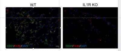
Immunohistochemistry-Frozen: F4/80 Antibody (BM8) [PE] [NBP2-22134] - Red: F4/80, Green: CD31/PECAM-1, Blue: DAPI+ nuclei. Frozen mouse lung tissues of WT B6 and IL1R--/- (knockout) mice were blocked with 1% BSA in PBS and were double stained with PE-conjugated F4/80 and Alex647-conjugated CD31/PECAM-1 antibody (MEC 7.46) for 2 hours at the room temperature. This image was submitted via a customer review.
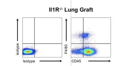
Flow Cytometry: F4/80 Antibody (BM8) [PE] [NBP2-22134] - Splenocytes isolated from a IL1R-/- mouse (IL1b receptor KO) were stained with F4/80 Antibody (BM8) and CD45-e450 (1:100 in FACS buffer) for 30 minutes. Cells were then washed and analyzed by flow cytometry. Left panel shows the isotype control. This image was submitted via a customer review.
Applications for F4/80 Antibody (BM8) [PE]
Application
Recommended Usage
Flow Cytometry
1:10-1:1000
Immunohistochemistry
1:10-1:500
Immunohistochemistry-Frozen
1:10-1:500
Immunohistochemistry-Paraffin
1:10-1:500
Western Blot
1:100-1:2000
Application Notes
This antibody works in Flow Cytometry, Immunohistochemistry-Frozen, Immunohistochemistry-Paraffin, Western Blot. The typical starting working dilution is 1:50. F4/80 is sensitive to 2-mercaptoethanol. The antibody requires proteinase K treatment of paraffin sections. IHC-P and Flow reported in a verified customer review.
Reviewed Applications
Read 2 reviews rated 5 using NBP2-22134 in the following applications:
Spectra Viewer
Plan Your Experiments
Use our spectra viewer to interactively plan your experiments, assessing multiplexing options. View the excitation and emission spectra for our fluorescent dye range and other commonly used dyes.
Spectra Viewer
Formulation, Preparation, and Storage
Purification
Protein A or G purified
Formulation
PBS and 1.0% BSA
Preservative
0.02% Sodium Azide
Concentration
Please see the vial label for concentration. If unlisted please contact technical services.
Shipping
The product is shipped with polar packs. Upon receipt, store it immediately at the temperature recommended below.
Stability & Storage
Store at 4C in the dark.
Background: F4/80
Depending on factors such as differentiation, activation, and location, the level of F4/80 expression can vary between cell-types (1,2). For example, peritoneal macrophages are subdivided based on morphology and expression with large peritoneal macrophages having high levels of F4/80 and small peritoneal macrophages having low F4/80 expression (1). Furthermore, splenic macrophages are classified based on location (red pulp, marginal zone, and white pulp) and F4/80 expression level varies within these populations as well; the red pulp macrophages have high levels of F4/80 and CD68 by comparison (1). Originally, the F4/80 monoclonal antibody was generated for a macrophage-specific surface antigen, however F4/80 staining is also observed in other hematopoietic cell populations, thus other immune cell markers should be used in conjunction with F4/80 to distinguish between different populations (1). Though the full extent of F4/80 ligands and biological functions remains elusive, studies have suggested that F4/80 is critical for macrophage-natural killer cell interaction and cytokine release (1,2). Additionally, a study of anterior chamber-associated immune deviation (ACAID) indicated a possible role for F4/80 in the generation of regulatory T cells (1,2).
References
1. Dos Anjos Cassado A. (2017). F4/80 as a Major Macrophage Marker: The Case of the Peritoneum and Spleen. Results and Problems in Cell Differentiation. https://doi.org/10.1007/978-3-319-54090-0_7
2. Hamann, J., Hsiao, C. C., Lee, C. S., Ravichandran, K. S., & Lin, H. H. (2016). Adhesion GPCRs as Modulators of Immune Cell Function. Handbook of Experimental Pharmacology. https://doi.org/10.1007/978-3-319-41523-9_15
Alternate Names
DD7A5-7, Gpf480, Ly71
Gene Symbol
Adgre1
Additional F4/80 Products
Product Documents for F4/80 Antibody (BM8) [PE]
Product Specific Notices for F4/80 Antibody (BM8) [PE]
This product is for research use only and is not approved for use in humans or in clinical diagnosis. Primary Antibodies are guaranteed for 1 year from date of receipt.
Loading...
Loading...
Loading...
Loading...
Loading...
Loading...