Rat Pure-Blot anti-Mouse IgG (H+L) Secondary Antibody (eB144) [DyLight 800]
Novus Biologicals, part of Bio-Techne | Catalog # NBP3-11663
Conjugate
Catalog #
Key Product Details
Species Reactivity
Mouse
Applications
Immunocytochemistry/ Immunofluorescence, Immunoprecipitation, Western Blot
Label
DyLight 800 (Excitation = 777 nm, Emission = 794 nm)
Antibody Source
Monoclonal Rat IgG Clone # eB144
Concentration
LYOPH mg/ml
Product Specifications
Immunogen
Mouse IgG
Clonality
Monoclonal
Host
Rat
Isotype
IgG
Description
This secondary antibody was prepared from tissue culture supernatant by Protein G affinity chromatography. Assay by Immunoelectrophoresis resulted in a single precipitin arc against Anti-Mouse Serum.
Store vial at 4C prior to restoration. For extended storage aliquot contents and freeze at -20C or below. Avoid cycles of freezing and thawing. Centrifuge product if not completely clear after standing at room temperature. This product is stable for several weeks at 4C as an undiluted liquid. Dilute only prior to immediate use.
Store vial at 4C prior to restoration. For extended storage aliquot contents and freeze at -20C or below. Avoid cycles of freezing and thawing. Centrifuge product if not completely clear after standing at room temperature. This product is stable for several weeks at 4C as an undiluted liquid. Dilute only prior to immediate use.
Scientific Data Images
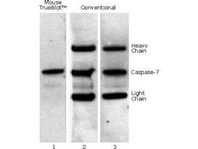
Western Blot: Rat Pure-Blot anti-Mouse IgG (H+L) Secondary Antibody (eB144) [DyLight 800] [NBP3-11663] - Caspase 7 was immunoprecipitated from 0.5 ml of 1x10e7 Jurkat cells/ml with 5 ug mouse anti-human Caspase 7. Precipitate from 1x10e6 cells was subjected to electrophoresis, transferred to an PVDF membrane, and Western blotted with anti-Caspase 7 using Rat Pure-Blot anti-Mouse IgG (H+L) Secondary Antibody (eB144) [DyLight 800] (Lane 1) or conventional HRP-conjugated anti-mouse antibody (Lane 2) - note the detection of the heavy and light chains of the immunoprecipitating antibody in Lane 2 but not in Lane 1. When Lane 1 is re-immunoblotted using conventional HRP-conjugated anti-mouse polyclonal antibody (Lane 3), the heavy and light chains are now detected, confirming that although the immunoprecipitating heavy and light chains are present, Rat Pure-Blot anti-Mouse IgG (H+L) Secondary Antibody (eB144) [DyLight 800] detects only native antibody and not denatured heavy and light chains.
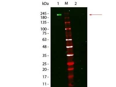
Western Blot: Rat Pure-Blot anti-Mouse IgG (H+L) Secondary Antibody (eB144) [DyLight 800] [NBP3-11663] - Lane 1: Mouse IgG, Non-reduced. Lane 2: Mouse IgG, Reduced. Load: 50 ng per lane. Primary antibody: none. Secondary antibody: Rat Pure-Blot anti-Mouse IgG (H+L) Secondary antibody (eB144) [DyLight 800] at 1:1,000 for 60 min at RT. Block for 30 min at RT. Predicted/Observed size: 160 kDa for Mouse IgG, Non-reduced. Migrates at slightly higher molecular weight than predicted.
![Rat Pure-Blot anti-Mouse IgG (H+L) Secondary Antibody (eB144) [DyLight 800]](https://resources.bio-techne.com/images/products/nbp3-11663_rat-pure-blot-anti-mouse-igg-h-l-secondary-antibody-eb144-dylight-800-255202313292311.jpg "Rat Pure-Blot anti-Mouse IgG (H+L) Secondary Antibody (eB144) [DyLight 800]")
Rat Pure-Blot anti-Mouse IgG (H+L) Secondary Antibody (eB144) [DyLight 800]
Western Blot of Fluorescent TrueBlot(R): Anti-Mouse Ig DyLight(TM) 800. Lane 1: Mouse IgG, Non-reduced. Lane 2: Mouse IgG, Reduced. Load: 50 ng per lane. Primary antibody: none. Secondary antibody: Fluorescent TrueBlot(R): Anti-Mouse Ig DyLight(TM) 800 at 1:1,000 for 60 min at RT. Block for 30 min at RT. Predicted/Observed size: 160 kDa for Mouse IgG, Non-reduced. Migrates at slightly higher molecular weight than predicted.Applications
Application
Recommended Usage
Immunocytochemistry/ Immunofluorescence
1:500 - 1:2500
Western Blot
1:1000
Application Notes
This antibody has been tested in western blot and immunoprecipitation and may also be used for detection in immunoassays that do not employ immunoprecipitation. This antibody is provided as a lyophilized powder. To conserve reagent, we recommend incubating the blots from minigels in sealed bags (removing as much air as possible) with minimal volume (2-3 mLs). If used conservatively at 2.5mls/blot will yield enough reagent for 40 blots. Note that there are three key procedural considerations: 1. Protein A or G beads may be used with the mouse, goat and sheep Pure-Blot secondaries, but not with the rabbit Pure-Blot secondary. Use of protein A or G beads with the rabbit Pure-Blot will result in contaminating bands. 2. Immunoprecipitate should be completely reduced. 3. Bovine Serum Albumin or blocking buffer for Fluorescent Western Blotting, at low concentrations, should be used as the blocking protein for the immunoblot. DO NOT USE BLOTTO or MILK. Note: To achieve best results when detecting mouse IgG1 subtypes, we recommend performing a dot blot or titration to determine the optimal dilution factor for your desired application. All recommended dilutions for listed applications are intended as an initial recommendation, specific conditions for each protein and antibody combination should be specifically optimized by the end user. Fluorescence technology is widely used to detect proteins. However, many common visible fluorophores often result in considerable background fluorescence in the visible range. Visible fluorophores are rarely used for membrane-based protein detection because of this high background. DyLight(TM) 800 and DyLight(TM) 680 antibody and reagent conjugates are specifically designed for protein detection methods that use longer-wavelength, near-infrared (IR) fluorophores to visualize proteins in western blotting and other applications. Very low background fluorescence in the IR range provides for a much higher signal-to-noise ratio than visible fluorophores. Detection levels in the picogram range on Western blots rival the sensitivity of chemiluminescence on film. DyLight(TM) 800 conjugates are optimized for the Odyssey(R) Infrared Imaging System developed by LI-COR. DyLight(TM) 800 conjugates are also suitable for immunofluorescence microscopy using commercially available excitation/emission filters in the 780nm/820nm range. Dual simultaneous labeling in western blots or microscopy is achieved when DyLight(TM) 800 conjugates are used in conjunction with DyLight(TM) 680 conjugates. DyLight(TM) 800 and DyLight(TM) 680 conjugates provide an ultra-sensitive and convenient alternative to standard chemiluminescent protein detection methods, as well as a valuable tool for multicolor imaging.
Formulation, Preparation, and Storage
Purification
Protein G purified
Reconstitution
Reconstitute with 100 ul deionized water (or equivalent).
Formulation
Lyophilized from 0.02 M Potassium Phosphate, 0.15 M Sodium Chloride, pH 7.2, 10 mg/ml Polyethylene Glycol (PEG-8000)
Preservative
0.01% Sodium Azide
Concentration
LYOPH mg/ml
Shipping
The product is shipped with polar packs. Upon receipt, store it immediately at the temperature recommended below.
Stability & Storage
Store lyophilized antibody at 4C. Aliquot reconstituted liquid and store at -20C. Avoid freeze-thaw cycles.
Background: IgG (H+L)
The 4 IgG subclasses, sharing 95% amino acid identity, include IgG1, IgG2, IgG3, and IgG4 for humans and IgG1, IgG2a, IgG2b, and IgG3 for mice. The relative abundance of each human subclass is 60% for IgG1, 32% for IgG2, 4% for IgG3, and 4% for IgG4. In an IgG deficiency, there may be a shortage of one or more subclasses (4).
References
1. Painter RH. (1998) Encyclopedia of Immunology (Second Edition). Elsevier. 1208-1211
2. Chapter 9 - Antibodies. (2012) Immunology for Pharmacy. Mosby 70-78
3. Schroeder H, Cavacini, L. (2010) Structure and Function of Immunoglobulins. J Allergy Clin Immunol. 125(2 0 2): S41-S52. PMID: 20176268
4. Vidarsson G, Dekkers G, Rispens T. (2014) IgG subclasses and allotypes: from structure to effector functions. Front Immunol. 5:520. PMID: 25368619
Alternate Names
IP Detection Reagent
Additional IgG (H+L) Products
Product Specific Notices
This product is for research use only and is not approved for use in humans or in clinical diagnosis. Secondary Antibodies are guaranteed for 1 year from date of receipt.
Loading...
Loading...
Loading...
Loading...
Loading...