
MortaLIVE™ Non-Toxic Dye
Tocris Bioscience, part of Bio-Techne | Catalog # 8936
Ready-to-use fluorescent live-dead cell stain for long term live cell imaging

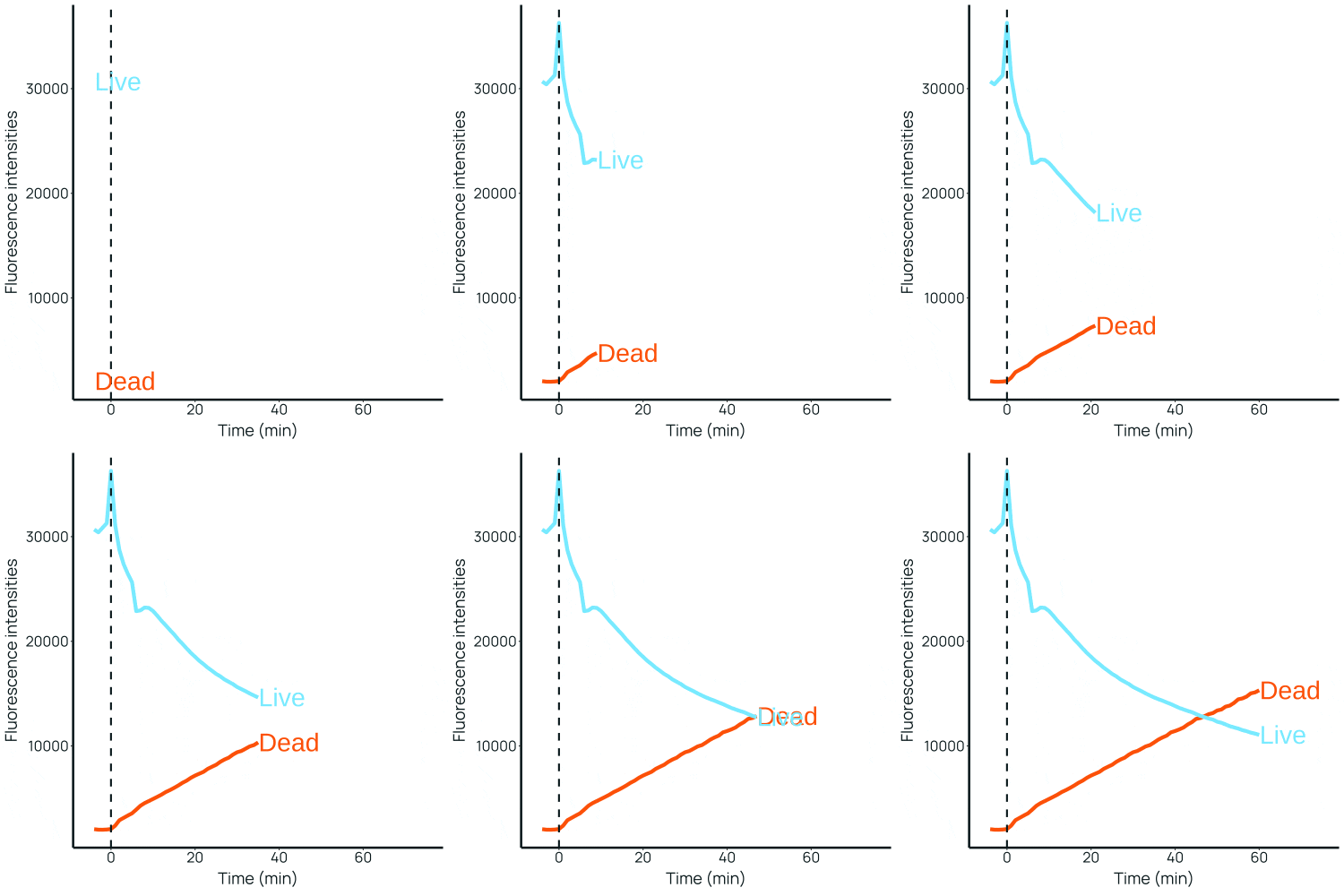
Key Product Details
Description
Ready-to-use fluorescent live-dead cell stain for long term live cell imaging
Product Description
MortaLIVE™ Non-Toxic Dye is a ready-to-use dual-color fluorescent live-dead cell stain for long term live-cell imaging. It can be used for toxicity assays and for tracking cell viability and cytotoxicity in real time by cell imaging. The live cell channel has an excitation maximum (λ) of 640 nm, while the dead cell channel has an excitation maximum (λ) of 488 nm. This dual-color format allows continuous, non toxic tracking of cell health over extended imaging periods without the need for endpoint staining. MortaLIVE™ Non-Toxic Dye is provided as 50 μL of a 1000x concentrate in DMSO.Please refer to this protocol for further information on how to use this product.
MortaLIVE™ is a trademark of Saguaro Biosciences.
Scientific Data Images for MortaLIVE™ Non-Toxic Dye

MortaLIVE™ Non-Toxic Dye Application.
Live-cell fluorescence imaging of heart organoids stained with MortaLIVE™ and NucleoLIVE™ for real-time quantification of cytotoxicity under hypoxia (top row, 4 h) or sham (bottom row, 3 h) conditions. Left column shows MortaLIVE™ staining alone; right column shows multiplexed staining with both MortaLIVE™ and NucleoLIVE™. Live cells are shown in purple, dead cells in green, and nuclei in red. Images acquired using a Zeiss Airyscan 880 confocal microscope (40× oil objective, 20 z-stacks at 1 µm step size).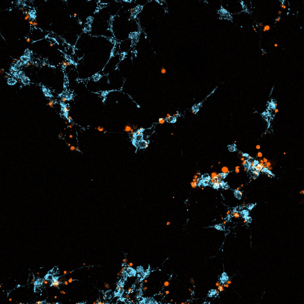
MortaLIVE™ Non-Toxic Dye Application.
Live-cell fluorescence imaging of MCF-7 cells stained with MortaLIVE™ and NucleoLIVE™ after 24 hours under compound-treated conditions. Nuclear channel not shown.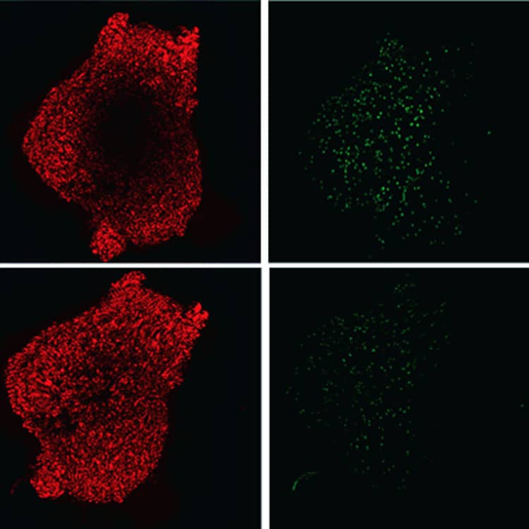
MortaLIVE™ Non-Toxic Dye Application.
Live-cell fluorescence imaging of heart organoids stained with MortaLIVE™ for real-time quantification of cytotoxicity under hypoxia (top row, 4 h) or sham (bottom row, 3 h) conditions. Images acquired using a Nikon-Crest spinning disk confocal microscope (20× air objective, 20 z-stacks at 5 µm step size). Left column shows live cells (red); right column shows dead cells (green).Licensing Information
Sold under license from Saguaro BiosciencesOptical Data
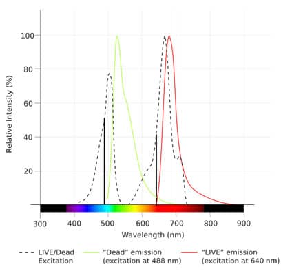
| λabs | 488 nm |
| λem | 640 nm |
| Cell Permeable | Yes |
Spectra Viewer
Plan Your Experiments
Use our spectra viewer to interactively plan your experiments, assessing multiplexing options. View the excitation and emission spectra for our fluorescent dye range and other commonly used dyes.
Spectra Viewer
Product Specifications
Storage
Store at -20°C
Purity
≥90% (HPLC)
The technical data provided above is for guidance only. For batch specific data refer to the Certificate of Analysis.
Solubility
| Solubility | Soluble in DMSO (supplied pre-dissolved in DMSO, 1000x) |
Calculators
Dilution Calculator
Reconstitution Calculator
Product Specific Notices for MortaLIVE™ Non-Toxic Dye
For research use only
Loading...
Loading...