
TGF-beta 2 Antibody
R&D Systems, part of Bio-Techne | Catalog # MAB612

Scientific images may be resized, cropped, or adjusted for brightness or contrast for presentation purposes. These updates are limited to presentation and do not affect the underlying scientific interpretation of the data. Where available, additional source data may be provided upon request.
Conjugate
Catalog #
Key Product Details
Species Reactivity
Validated:
Multi-Species
Cited:
Human, Mouse, Xenograft
Applications
Validated:
Immunohistochemistry, Western Blot, ELISA Capture (Matched Antibody Pair)
Cited:
Western Blot, Neutralization, Immunocytochemistry, ELISA Capture, ELISA Development
Label
Unconjugated
Antibody Source
Monoclonal Mouse IgG2B Clone # 8607
Product Specifications
Immunogen
Porcine TGF-beta 1.2
Specificity
Detects TGF-beta 2 from multiple species in ELISAs and Western blots. In ELISAs, no cross-reactivity or interfereence with recombinant human (rh) TGF-alpha, recombinant porcineTGF-beta 1, rhTGF-beta 1, recombinant canine TGF-beta 3, recombinant amphibian TGF-beta 5, rhTGF-beta RII, or rhTGF-beta RIII is observed.
Clonality
Monoclonal
Host
Mouse
Isotype
IgG2B
Scientific Data Images for TGF-beta 2 Antibody
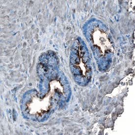
TGF‑ beta2 in Human Prostate Cancer.
TGF-beta 2 was detected in immersion fixed paraffin-embedded sections of human prostate cancer tissue using Mouse Anti-TGF-beta 2 Monoclonal Antibody (Catalog # MAB612) at 15 µg/mL overnight at 4 °C. Tissue was stained using the Anti-Mouse HRP-DAB Cell & Tissue Staining Kit (brown; Catalog # CTS002) and counterstained with hematoxylin (blue). Specific staining was localized to cytoplasm in cancer cells. View our protocol for Chromogenic IHC Staining of Paraffin-embedded Tissue Sections.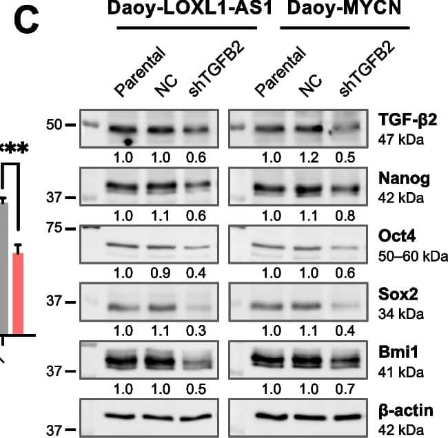
Detection of TGF-beta 2 by Western Blot
Effects of transforming growth factor (TGF)-beta 2 silencing on metastatic phenotypes of sonic-hedgehog medulloblastoma cells with high LOXL1-AS1 expression. A RT-qPCR analysis of TGF-beta 2 expression in Daoy-LOXL1-AS1 and Daoy-MYCN cells. B RT-qPCR analysis of four stemness markers in Daoy-LOXL1-AS1 and Daoy-MYCN cells relative to the parental group. C Western blot analysis of four stemness markers in Daoy-LOXL1-AS1 and Daoy-MYCN cells. Quantification values below each band are relative to the parental group. D, G Sphere-formation assay of (D) Daoy-LOXL1-AS1 and (G) Daoy-MYCN cells, including microscopic images (left images), and the number and sizes of spheres of ≥150 μm in diameter (right bar chart). Scale bars, 500 μm in 4× and 100 μm in 20× magnification. E, H Wound-healing assay of (E) Daoy-LOXL1-AS1 and (H) Daoy-MYCN cells. F, I Transwell migration and invasion assays of (F) Daoy-LOXL1-AS1 and (I) Daoy-MYCN cells including quantification of cell migration (left bar chart) and invasion (right bar chart). Quantitative data are presented as mean ± SD of replicates from representative of three independent experiments. NC, negative control; ns, non-significant. * p < 0.05, ** p < 0.01, *** p < 0.001 Image collected and cropped by CiteAb from the following open publication (https://jeccr.biomedcentral.com/articles/10.1186/s13046-024-03057-0), licensed under a CC-BY license. Not internally tested by R&D Systems.
Human TGF-beta 2 ELISA Standard Curve
Recombinant Human TGF-beta 2 (Catalog # 302-B2) was serially diluted and captured by Mouse Anti-TGF-beta 2 Monoclonal Antibody (Catalog # MAB612) coated on a Clear Polystyrene Microplate (Catalog # DY990). Goat Anti-TGF-beta 2 Antigen Affinity-purified Polyclonal Antibody (Catalog # AF-302-NA) was biotinylated and incubated with the protein captured on the plate. Detection of the standard curve was achieved by incubating Streptavidin-HRP (Catalog # DY998)Applications for TGF-beta 2 Antibody
Application
Recommended Usage
Immunohistochemistry
8-25 µg/mL
Sample: Immersion fixed paraffin-embedded sections of human prostate cancer tissue
Sample: Immersion fixed paraffin-embedded sections of human prostate cancer tissue
Western Blot
1 µg/mL
Sample: Recombinant Human TGF-beta 2 (Catalog # 302-B2)
under non-reducing conditions only
Sample: Recombinant Human TGF-beta 2 (Catalog # 302-B2)
under non-reducing conditions only
Human TGF-beta 2 Sandwich Immunoassay
Please Note: Optimal dilutions of this antibody should be experimentally determined.
Reviewed Applications
Read 4 reviews rated 5 using MAB612 in the following applications:
Formulation, Preparation, and Storage
Purification
Protein A or G purified from ascites
Reconstitution
Reconstitute at 0.5 mg/mL in sterile PBS. For liquid material, refer to CoA for concentration.
Formulation
Lyophilized from a 0.2 μm filtered solution in PBS with Trehalose. *Small pack size (SP) is supplied either lyophilized or as a 0.2 µm filtered solution in PBS.
Shipping
Lyophilized product is shipped at ambient temperature. Liquid small pack size (-SP) is shipped with polar packs. Upon receipt, store immediately at the temperature recommended below.
Stability & Storage
Use a manual defrost freezer and avoid repeated freeze-thaw cycles.
- 12 months from date of receipt, -20 to -70 °C as supplied.
- 1 month, 2 to 8 °C under sterile conditions after reconstitution.
- 6 months, -20 to -70 °C under sterile conditions after reconstitution.
Background: TGF-beta 2
Long Name
Transforming Growth Factor beta 2
Alternate Names
TGFB2, TGFbeta 2
Gene Symbol
TGFB2
Additional TGF-beta 2 Products
Product Documents for TGF-beta 2 Antibody
Product Specific Notices for TGF-beta 2 Antibody
For research use only
Loading...
Loading...
Loading...
Loading...
Loading...