
Rat ICAM-1/CD54 Biotinylated Antibody
R&D Systems, part of Bio-Techne | Catalog # BAF583

Scientific images may be resized, cropped, or adjusted for brightness or contrast for presentation purposes. These updates are limited to presentation and do not affect the underlying scientific interpretation of the data. Where available, additional source data may be provided upon request.
Conjugate
Catalog #
Key Product Details
Species Reactivity
Rat
Applications
Western Blot, ELISA Detection (Matched Antibody Pair)
Label
Biotin
Antibody Source
Polyclonal Goat IgG
Product Specifications
Immunogen
Mouse myeloma cell line NS0-derived recombinant rat ICAM‑1/CD54
Gln28-Thr493
Accession # Q00238
Gln28-Thr493
Accession # Q00238
Specificity
Detects rat ICAM‑1/CD54 in ELISAs and Western blots. In sandwich immunoassays, less than 0.1% cross-reactivity with recombinant mouse (rm) ICAM-1, recombinant human (rh) ICAM-1, rhICAM-3, rmICAM-5, and rmICAM-2 is observed.
Clonality
Polyclonal
Host
Goat
Isotype
IgG
Scientific Data Images for Rat ICAM-1/CD54 Biotinylated Antibody
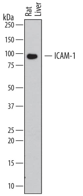
Detection of Rat ICAM‑1/CD54 by Western Blot.
Western blot shows lysates of rat liver tissue. PVDF membrane was probed with 0.2 µg/mL of Goat Anti-Rat ICAM-1/CD54 Biotinylated Antigen Affinity-purified Polyclonal Antibody (Catalog # BAF583) followed by Streptavidin-HRP (Catalog # DY998). A specific band was detected for ICAM-1/CD54 at approximately 90 kDa (as indicated). This experiment was conducted under reducing conditions and using Immunoblot Buffer Group 1.
Rat ICAM-1 / CD54 ELISA Standard Curve
Recombinant Rat ICAM-1/CD54 Fc Chimera (Catalog # 583-IC) was serially diluted and captured by Mouse Anti-Rat ICAM-1/CD54 Monoclonal Antibody (Catalog # MAB5832) coated on a Clear Polystyrene Microplate (Catalog # DY990). Goat Anti-Rat ICAM-1/CD54 Antigen Affinity-purified Polyclonal Antibody (Catalog # AF583) was biotinylated and incubated with the protein captured on the plate. Detection of the standard curve was achieved by incubating Streptavidin-HRP (Catalog # DY998)Applications for Rat ICAM-1/CD54 Biotinylated Antibody
Application
Recommended Usage
Western Blot
0.2 µg/mL
Sample: Rat liver tissue
Sample: Rat liver tissue
Rat ICAM-1/CD54 Sandwich Immunoassay
Please Note: Optimal dilutions of this antibody should be experimentally determined.
Formulation, Preparation, and Storage
Purification
Antigen Affinity-purified
Reconstitution
Reconstitute at 0.2 mg/mL in sterile PBS.
Formulation
Lyophilized from a 0.2 μm filtered solution in PBS with BSA as a carrier protein.
Shipping
The product is shipped at ambient temperature. Upon receipt, store it immediately at the temperature recommended below.
Stability & Storage
Use a manual defrost freezer and avoid repeated freeze-thaw cycles.
- 12 months from date of receipt, -20 to -70 °C as supplied.
- 1 month, 2 to 8 °C under sterile conditions after reconstitution.
- 6 months, -20 to -70 °C under sterile conditions after reconstitution.
Background: ICAM-1/CD54
Intercellular Adhesion Molecule-1 (ICAM-1, CD54), binds the leukocyte integrins LFA-1 and Mac-1. ICAM-1 expression is weak on leukocytes, epithelial and resting endothelial cells, as well as some other cell types, but expression can be stimulated by IFN-gamma, TNF-alpha, IL-1 beta, and lipolysaccharide (LPS). Rat and human ICAM-1 share approximately 52% amino acid identity. Rat and mouse ICAM-1 share approximately 52% amino acid identity.
Soluble ICAM-1 is found in a biologically active form in serum, probably as a result of proteolytic cleavage from the cell surface, and is elevated in patients with various inflammatory syndromes such as septic shock, leukocyte adhesion deficiency syndrome (LAD), cancer, and transplantation.
References
- Pigott, R. and C. Power (1993) in The Adhesion Molecule Facts Book, p. 74. Academic Press.
- Siu, G. et al. (1989) J. Immunol. 143:3813.
- Ballantyne, C.M. et al. (1989) Nuc. Acid. Res. 17:5853.
Long Name
Intercellular Adhesion Molecule 1
Alternate Names
CD54, ICAM1
Gene Symbol
ICAM1
UniProt
Additional ICAM-1/CD54 Products
Product Documents for Rat ICAM-1/CD54 Biotinylated Antibody
Product Specific Notices for Rat ICAM-1/CD54 Biotinylated Antibody
For research use only
Loading...
Loading...
Loading...
Loading...
Loading...