Mouse Syndecan-2/CD362 Antibody
R&D Systems, part of Bio-Techne | Catalog # AF6585


Key Product Details
Species Reactivity
Validated:
Cited:
Applications
Validated:
Cited:
Label
Antibody Source
Product Specifications
Immunogen
Glu19-Phe141
Accession # P43407
Specificity
Clonality
Host
Isotype
Scientific Data Images for Mouse Syndecan-2/CD362 Antibody
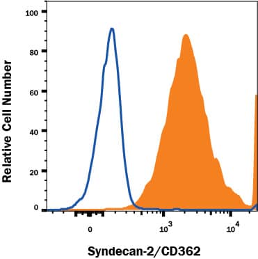
Detection of Syndecan-2/CD362 in NIH-3T3 Mouse Cell Line by Flow Cytometry.
NIH-3T3 mouse cell line was stained with Sheep Anti-Mouse Syndecan-2/CD362 Antigen Affinity-purified Polyclonal Antibody (Catalog # AF6585, filled histogram) or control antibody (Catalog # 5-001-A, open histogram), followed by Allophycocyanin-conjugated Anti-Sheep IgG Secondary Antibody (Catalog # F0127). View our protocol for Staining Membrane-associated Proteins.Applications for Mouse Syndecan-2/CD362 Antibody
CyTOF-ready
Flow Cytometry
Sample: NIH-3T3 mouse cell line
Formulation, Preparation, and Storage
Purification
Reconstitution
Formulation
*Small pack size (-SP) is supplied either lyophilized or as a 0.2 µm filtered solution in PBS.
Shipping
Stability & Storage
- 12 months from date of receipt, -20 to -70 °C as supplied.
- 1 month, 2 to 8 °C under sterile conditions after reconstitution.
- 6 months, -20 to -70 °C under sterile conditions after reconstitution.
Background: Syndecan-2/CD362
Syndecan-2, previously known as fibroglycan or heparan sulfate proteoglycan, is a member of the syndecan family of type 1 transmembrane proteins capable of carrying heparan sulfate (HS) and chondroitin sulfate glycosaminoglycans. The four vertebrate syndecans show conserved cytoplasmic domains and divergent extracellular portions (except for GAG attachment sites). Among the Syndecans, Syndecan-2 is most similar to Syndecan-4 (1‑3). Mouse Syndecan-2 is synthesized as a 202 amino acid (aa) core protein with an 18 aa signal sequence, a 127 aa extracellular domain (ECD), a 25 aa transmembrane region and a 32 aa cytoplasmic tail (4). The ECD of mouse Syndecan-2 contains three closely-spaced consensus Ser-Gly sequences for the attachment of HS side chains. It shares 76%, 86%, 74% and 72% aa identity with the ECD of human, rat, porcine and bovincoe Syndecan-2, respectively. The cytoplasmic tail has both serine and tyrosine phosphorylation sites. Addition of 20 ‑ 80 disaccharides per side chain adds considerably to the size of the 22 kDa core protein. Non-covalent homodimerization of Syndecan-2, or heterodimerization with Syndecan-4, is dependent on the transmembrane domain (5, 6). Syndecan-2 is expressed in cells of mesenchymal origin, neuronal and epithelial cells, and is the predominant syndecan expressed during embryonic development. Expression is up‑regulated in several cancer cell lines (7). After induction in macrophages by inflammatory mediators, Syndecan-2 selectively binds FGF basic, VEGF and EGF (8). Syndecan-2 expressed on human primary osteoblasts binds GM‑CSF and may function as a co-receptor (9). Activated endothelial cell Syndecan-2 specifically binds IL-8 and may participate in promoting neutrophil extravasation by forming a chemotactic IL-8 gradient (10). Typically, cytokine, chemokine and extracellular matrix protein binding occurs through interaction with HS side chains, but the Syndecan-2 extracellular domain can bind TGF-beta directly via protein-protein interaction (11).
References
- Tkachenko, E. et al. (2005) Circ. Res. 96:488.
- Oh, E.-S, and J. R. Couchman (2004) Mol. Cells 17:181.
- Essner, J. J. et al. (2006) Int. J. Biochem. Cell Biol. 38:152.
- Marynen, P. et al. (1989) J. Biol. Chem. 264:7017.
- Choi, S. et al. (2005) J. Biol. Chem. 280:42573.
- Dews, I.C. and K.R. MacKenzie (2007) Proc. Natl. Acad. Sci. USA 104:20782.
- Park, H. et al. (2002) J. Biol. Chem. 277:29730.
- Clasper, S. et al. (1999) J. Biol. Chem. 274:24113.
- Modrowski, D. et al. (2000) J. Biol. Chem. 275:9178.
- Halden, Y. et al. (2004) Biochem. J. 377:533.
- Chen, L. et al. (2004) J. Biol. Chem. 279:15715.
Alternate Names
Gene Symbol
UniProt
Additional Syndecan-2/CD362 Products
Product Documents for Mouse Syndecan-2/CD362 Antibody
Product Specific Notices for Mouse Syndecan-2/CD362 Antibody
For research use only