
Mouse Syndecan-1/CD138 Antibody
R&D Systems, part of Bio-Techne | Catalog # AF3190

Scientific images may be resized, cropped, or adjusted for brightness or contrast for presentation purposes. These updates are limited to presentation and do not affect the underlying scientific interpretation of the data. Where available, additional source data may be provided upon request.
Conjugate
Catalog #
Key Product Details
Species Reactivity
Validated:
Mouse
Cited:
Human, Mouse
Applications
Validated:
Immunohistochemistry, Western Blot, Flow Cytometry, Immunocytochemistry, CyTOF-ready
Cited:
Immunohistochemistry, Western Blot, Flow Cytometry, Immunocytochemistry
Label
Unconjugated
Antibody Source
Polyclonal Goat IgG
Product Specifications
Immunogen
Mouse myeloma cell line NS0-derived recombinant mouse Syndecan‑1/CD138 isoform 1
Gln18-Glu252
Accession # P18828
Gln18-Glu252
Accession # P18828
Specificity
Detects mouse Syndecan‑1/CD138 in direct ELISAs and Western blots. In Western blots, approximately 5% cross-reactivity with recombinant human (rh) Syndecan-1 is observed and less than 1% cross-reactivity with rhSyndecan-2, recombinant mouse (rm) Syndecan-3 and rmSyndecan-4 is observed.
Clonality
Polyclonal
Host
Goat
Isotype
IgG
Scientific Data Images for Mouse Syndecan-1/CD138 Antibody
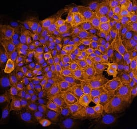
Syndecan‑1/CD138 in NMuMG Mouse Cell Line.
Syndecan-1/CD138 was detected in immersion fixed NMuMG mouse mammary gland epithelial cell line using Mouse Syndecan-1/CD138 Antigen Affinity-purified Polyclonal Antibody (Catalog # AF3190) at 10 µg/mL for 3 hours at room temperature. Cells were stained using the NorthernLights™ 557-conjugated Anti-Goat IgG Secondary Antibody (yellow; Catalog # NL001) and counterstained with DAPI (blue). Specific staining was localized to cell surfaces and cytoplasm. View our protocol for Fluorescent ICC Staining of Cells on Coverslips.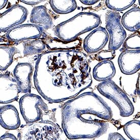
Syndecan‑1/CD138 in Mouse Kidney.
Syndecan-1/CD138 was detected in perfusion fixed frozen sections of mouse kidney using Goat Anti-Mouse Syndecan-1/CD138 Antigen Affinity-purified Polyclonal Antibody (Catalog # AF3190) at 3 µg/mL for 1 hour at room temperature followed by incubation with the Anti-Goat IgG VisUCyte™ HRP Polymer Antibody (Catalog # VC004). Tissue was stained using DAB (brown) and counterstained with hematoxylin (blue). Specific staining was localized to glomeruli and convoluted tubules. View our protocol for IHC Staining with VisUCyte HRP Polymer Detection Reagents.
Detection of Syndecan-1 by Immunocytochemistry/ Immunofluorescence
Chemotherapy inhibits cytotoxic T lymphocyte activity. A) Gene set enrichment analysis (GSEA) of public RNA‐seq datasets (GSE161801 (n = 30) and GSE223060 (n = 41)) of the immune‐response‐gene‐related signatures between newly diagnosed patients, undergoing treatment and those in relapse. NES, normalized enrichment score. B) Representative two patient's UMAP plot of the expression of CD3D, CD8, CD69, and IFNG in a public scRNA‐seq data (GSE161801) (n = 30). UMAP: uniform manifold approximation and projection. C,D) Pathway enrichment analysis of public RNA‐seq datasets of the myeloma cells treated with or without melphalan (GSE178292) or bortezomib (GSE171837). E) Immunofluorescent staining of healthy and myeloma patients bone marrow samples before or after chemotherapy with 4',6‐diamidino‐2‐phenylindole dihydrochloride (DAPI) and antibodies against CD138 (myeloma cell marker) or granzyme B (GZB, activity of T cell) (n = 5). Scale bar, 10 µm. F) Box plots showing the relative mean fluorescence intensity (MFI) of CD138 (left panel) or granzyme B (right panel) in the biopsy segment (n = 5). G) The relative expression of TIM3, TOX, TIGIT, PD‐1, or LAG3 in CD8+ T cells isolated from the bone marrow aspirates of another 20 myeloma patients before or after chemotherapy. Image collected and cropped by CiteAb from the following open publication (https://pubmed.ncbi.nlm.nih.gov/40135791), licensed under a CC-BY license. Not internally tested by R&D Systems.Applications for Mouse Syndecan-1/CD138 Antibody
Application
Recommended Usage
CyTOF-ready
Ready to be labeled using established conjugation methods. No BSA or other carrier proteins that could interfere with conjugation.
Flow Cytometry
2.5 µg/106 cells
Sample: T1165 mouse plasmacytoma cell line
Sample: T1165 mouse plasmacytoma cell line
Immunocytochemistry
5-15 µg/mL
Sample: Immersion fixed NmuMG mouse normal mammary epithelial cell line
Sample: Immersion fixed NmuMG mouse normal mammary epithelial cell line
Immunohistochemistry
3-15 µg/mL
Sample: Perfusion fixed frozen sections of mouse kidney
Sample: Perfusion fixed frozen sections of mouse kidney
Western Blot
0.1 µg/mL
Sample: Recombinant Mouse Syndecan-1/CD138 (Catalog # 3190-SD)
Sample: Recombinant Mouse Syndecan-1/CD138 (Catalog # 3190-SD)
Reviewed Applications
Read 1 review rated 5 using AF3190 in the following applications:
Formulation, Preparation, and Storage
Purification
Antigen Affinity-purified
Reconstitution
Reconstitute at 0.2 mg/mL in sterile PBS. For liquid material, refer to CoA for concentration.
Formulation
Lyophilized from a 0.2 μm filtered solution in PBS with Trehalose. *Small pack size (SP) is supplied either lyophilized or as a 0.2 µm filtered solution in PBS.
Shipping
Lyophilized product is shipped at ambient temperature. Liquid small pack size (-SP) is shipped with polar packs. Upon receipt, store immediately at the temperature recommended below.
Stability & Storage
Use a manual defrost freezer and avoid repeated freeze-thaw cycles.
- 12 months from date of receipt, -20 to -70 °C as supplied.
- 1 month, 2 to 8 °C under sterile conditions after reconstitution.
- 6 months, -20 to -70 °C under sterile conditions after reconstitution.
Background: Syndecan-1/CD138
Syndecan-1 (also known as CD138) is a variably glycosylated, dimeric, type I transmembrane (TM) protein that belongs to the Syndecan family. It is synthesized as a 311 amino acid (aa) precursor that contains a 17 aa signal sequence, a 235 aa extracellular domain (ECD), a 25 aa TM segment, and a 34 aa cytoplasmic region. The ECD shows various degrees of heparan sulfate and chondroitin sulfate modification, leading to native molecular weights for Syndecan-1 of 120‑200 kDa. Proteolytic cleavage of the membrane-bound ECD yields soluble forms of approximately the same molecular weight. Syndecan-1 is an epithelial cell Syndecan involved in Wnt and chemokine signaling. The amino acid sequence of mouse Syndecan-1 ECD shares 71% identity with that of the ECD of both rat and human Syndecan-1.
Alternate Names
CD138, SDC1, Syndecan1
Gene Symbol
SDC1
UniProt
Additional Syndecan-1/CD138 Products
Product Documents for Mouse Syndecan-1/CD138 Antibody
Product Specific Notices for Mouse Syndecan-1/CD138 Antibody
For research use only
Loading...
Loading...
Loading...
Loading...
Loading...
Loading...