
Mouse CD8 alpha Antibody
R&D Systems, part of Bio-Techne | Catalog # MAB11715

Conjugate
Catalog #
Key Product Details
Species Reactivity
Mouse
Applications
Multiplex Immunofluorescence, Immunohistochemistry, COMET
Label
Unconjugated
Antibody Source
Monoclonal Rat IgG2A Clone # 1104516
Product Specifications
Immunogen
Mouse myeloma cell line, NS0-derived mouse CD8
Lys28-Tyr196
Accession # P01731
Lys28-Tyr196
Accession # P01731
Specificity
Detects recombinant mouse CD8 alpha protein in Direct ELISA.
Clonality
Monoclonal
Host
Rat
Isotype
IgG2A
Scientific Data Images for Mouse CD8 alpha Antibody
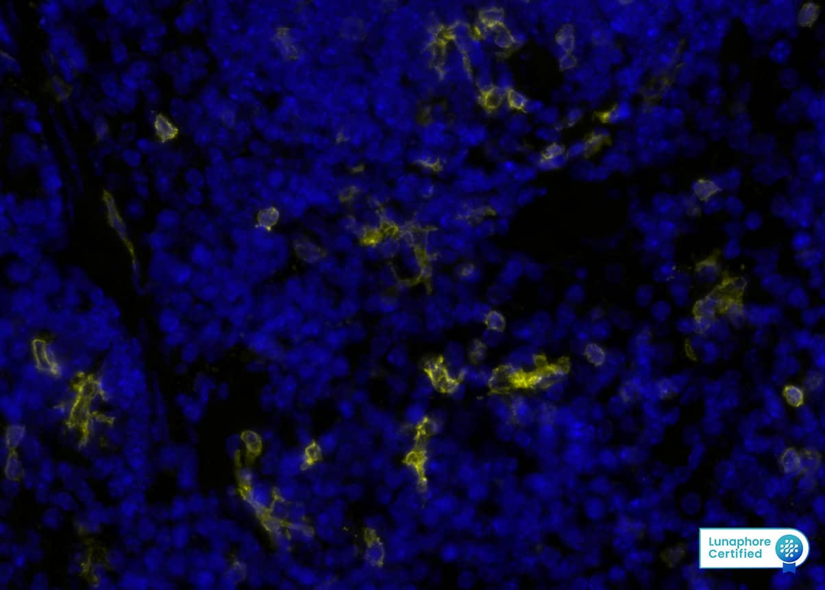
Detection of CD8a in Mouse spleen via seqIF™ staining on COMET™
CD8a was detected in perfusion fixed paraffin-embedded sections of mouse spleen using Rat Anti-Mouse CD8a, Monoclonal Antibody (Catalog #MAB11715) at 5ug/mL at 37 ° Celsius for 4 minutes. Before incubation with the primary antibody, tissue underwent an all-in-one dewaxing and antigen retrieval preprocessing using PreTreatment Module (PT Module) and Dewax and HIER Buffer H (pH 9; Epredia Catalog # TA-999-DHBH). Tissue was stained using the Alexa Fluor™ 647 Goat anti-Rat IgG Secondary Antibody at 1:200 at 37 ° Celsius for 2 minutes. (Yellow; Lunaphore Catalog # DR647RT) and counterstained with DAPI (blue; Lunaphore Catalog # DR100). Specific staining was localized to the membrane. Protocol available in COMET™ Panel Builder.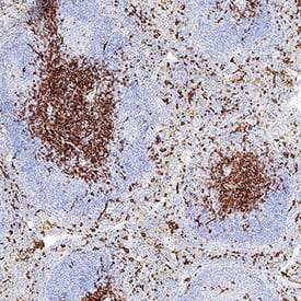
Detection of CD8 alpha in Mouse Spleen.
CD8 alpha was detected in perfusion fixed paraffin-embedded sections of mouse spleen using Rat Anti-Mouse CD8 alpha Monoclonal Antibody (Catalog # MAB11715) at 5 µg/ml overnight at 4 °C. Before incubation with the primary antibody, tissue was subjected to heat-induced epitope retrieval using VisUCyte Antigen Retrieval Reagent-Basic (Catalog # VCTS021). Tissue was stained using the HRP-conjugated Anti-Rat IgG Secondary Antibody (Catalog # HAF005) and counterstained with hematoxylin (blue). Specific staining was localized to the membrane. View our protocol for Chromogenic IHC Staining of Paraffin-embedded Tissue Sections.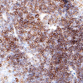
Detection of CD8 alpha in Mouse Thymus.
CD8 alpha was detected in perfusion fixed paraffin-embedded sections of mouse thymus using Rat Anti-Mouse CD8 alpha Monoclonal Antibody (Catalog # MAB11715) at 5 µg/ml overnight at 4 °C. Before incubation with the primary antibody, tissue was subjected to heat-induced epitope retrieval using VisUCyte Antigen Retrieval Reagent-Basic (Catalog # VCTS021). Tissue was stained using the HRP-conjugated Anti-Rat IgG Secondary Antibody (Catalog # HAF005) and counterstained with hematoxylin (blue). Specific staining was localized to the membrane. View our protocol for Chromogenic IHC Staining of Paraffin-embedded Tissue Sections.Applications for Mouse CD8 alpha Antibody
Application
Recommended Usage
COMET
Immunohistochemistry
3-25 µg/mL
Sample: Perfusion fixed paraffin-embedded sections of mouse spleen and thymus
Sample: Perfusion fixed paraffin-embedded sections of mouse spleen and thymus
Multiplex Immunofluorescence
5 µg/mL
Sample: Perfusion fixed paraffin-embedded sections of mouse spleen
Sample: Perfusion fixed paraffin-embedded sections of mouse spleen
Formulation, Preparation, and Storage
Purification
Protein A or G purified from hybridoma culture supernatant
Reconstitution
Reconstitute lyophilized material at 0.2 mg/ml in sterile PBS. For liquid material, refer to CoA for concentration.
Formulation
Lyophilized from a 0.2 μm filtered solution in PBS with Trehalose.
Shipping
Lyophilized product is shipped at ambient temperature. Liquid small pack size (-SP) is shipped with polar packs. Upon receipt, store immediately at the temperature recommended below.
Stability & Storage
Use a manual defrost freezer and avoid repeated freeze-thaw cycles.
- 12 months from date of receipt, -20 to -70 °C as supplied.
- 1 month, 2 to 8 °C under sterile conditions after reconstitution.
- 6 months, -20 to -70 °C under sterile conditions after reconstitution.
Background: CD8
CD8, also known as Ly-2, is a heterodimeric glycoprotein consisting of an alpha and beta chain. It is expressed on cytolytic T cells and functions in conjunction with the T cell receptor in the recognition of MHC/peptide complexes. Mouse CD8 (containing an alpha/Ly-2 or alpha′/Lyt-2 chain) is an antigen co‑receptor on the T cell surface which interacts with MHC I molecules on antigen presenting cells (1). CD8 alpha beta heterodimer is expressed on a subpopulation of mature T cells (2, 3). CD8 alpha, without CD8 beta, has been detected on subsets of gamma delta TCR-bearing T cells (4), intestinal intrathymic lymphocytes (5, 6) and dendritic cells (7, 8).
References
- Bierer, B.E. et al. (1989) Annu. Rev. Immunol. 7:579.
- Ledbetter, J.A. et al. (1980) J. Exp. Med. 152:280.
- Hayakawa, K. et al. (1994) Science 263:1131.
- MacDonald, H.R. et al. (1990) Eur. J. Immunol. 20:927.
- Rocha, B. et al. (1992) Immunol. Today 13:449.
- Wang, J. and J.R. Klein (1994) Science 265:1860.
- Vermec, D. et al. (1992) J. Exp. Med. 176:47.
- Suss, G. and K. Shortman (1996) J. Exp. Med. 183:1789.
Alternate Names
CD8, CD8A
Entrez Gene IDs
Gene Symbol
CD8A
UniProt
Additional CD8 Products
Product Documents for Mouse CD8 alpha Antibody
Product Specific Notices for Mouse CD8 alpha Antibody
For research use only
Loading...
Loading...
Loading...
Loading...
Loading...